등 근막이완요법이 30–60대 성인 여성의 발 형태에 미치는 영향
Effects of Back Fascia Relaxation Therapy on Foot Shape of Women in their 30–60s
背筋膜松弛疗法对30–60岁成年女性足部形态的影响
Article information
Abstract
목적
본 연구는 등 근막이완요법이 30–60대 성인 여성들의 발 형태에 영향을 미치는지를 살펴보고 관리 빈도와 연령에 따른 차이가 있는지도 알아보고자 하였다.
방법
부산에 거주하는 30–60대의 건강한 여성 34명을 관리 빈도(주1회, 주2회)에 따라 분류하고, 연령에 따라 30–40대와 50–60대로 구분하여 등 근막이완요법을 6회씩 진행하였다. 실험 전후에 족문을 찍어서 발의 형태 변화를 확인하였다. 족문을 통해 족장Ⅰ, 족장Ⅱ, 족폭, 종골폭, 종골각, 아치각, 모지각, 족선각, 외번각을 구하였다 자.료분석은 Statistical Package for the Social Sciences (SPSS) 20.0을 이용하여 독립 t 검정과 기술통계, 반복측정 이원분산분석을 실시하였다.
결과
등근막이완요법을 진행한 후 족문에 변화가 나타났다. 종골폭(p<0.01; p<0.001), 아치각(p<0.001; p<0.01), 모지각(p<0.01; p<0.01)은 왼발, 오른발 모두에서 측정 시기에 따라 통계적 유의성이 나타났고, 족장Ⅱ (p<0.05), 종골각(p<0.05), 외번각(p<0.01)은 오른발에서 유의미한 차이가 보였다. 그러나 족장Ⅰ, 족폭, 족선각에서는 유의한 변화가 보이지 않았다. 또한 연령과 관리 빈도에 따른 효과도 나타나지 않아 성인 여성들에게 등 근막이완요법이 연령과 관리 빈도에 관계없이 효과가 있음을 알 수 있었다.
결론
등 근막이완요법은 성인 여성의 발 형태에 변화를 주며 특히, 발의 척추 반사구와 관련성이 높음을 알 수 있었다.
Trans Abstract
Purpose
In this study, we investigated whether back fascia relaxation therapy could affect the foot shape of adult women in their 30–60s and also checked whether there were differences according to the frequency of back fascia therapy or participant age.
Methods
Based on the frequency of therapy (once a week vs. twice a week) and age (30–40s vs. 50–60s), 34 healthy women in their 30–60s living in Busan were divided into groups and underwent back fascia relaxation therapy six times. Footprinting was performed before and after the experiment (i.e., therapy) to confirm changes in the foot shape. Through footprinting, foot length I, foot length II, foot width, calcaneus width, heel angle, arch angle, hallux angle, toe angle, and eversion angle were measured. To analyze data, independent t-test, descriptive statistics, and two-way analysis of variance (ANOVA) of repeated measurements were performed using Statistical Package for the Social Sciences (SPSS) 20.0.
Results
The back fascia relaxation therapy affected the footprint appearance. The calcaneus width (p <0.01, p <0.001), arch angle (p <0.001, p <0.01), and hallux angle (p <0.01, p <0.01) were significantly different in both feet according to the measuring time. Further, foot length II (p <0.05), heel angle (p <0.05), and eversion angle (p <0.01) were significantly different in the right foot. However, there was no significant change in foot length I, foot width, or toe angle. Moreover, these results were consistent regardless of age or frequency of therapy.
Conclusion
Back fascia relaxation therapy changed the foot shape of adult women and was found to be highly relevant to the spinal reflex of the foot.
Trans Abstract
目的
探讨背筋膜松弛疗法是否会影响30–60岁成年女性的足部形态,并探讨根据背筋膜治疗频率和参与者年龄是否具有差异。
方法
对居住在釜山的30–60岁的健康女性,根据治疗频率(每周一次,每周两次)和年龄 分组,进行六次背筋膜松弛治疗。足迹是在实验前和实验后进行,并确认足部形态变化。通过足迹,测定脚长 I、脚长II、脚宽、跟骨宽度、倾角、拱角、拇角、束角以及外翻角。采用Statistical Package for the Social Sciences(SPSS)20.0进行独立样本t检验,描述性统计和重复测量的双向方差分析。
结果
背筋膜松弛疗 法进行后,足迹产生变化。根据测量时间,跟骨宽度(p <0.01,p <0.001),拱角(p <0.001,p <0.01) 以及拇角(p <0.01, p <0.01)在左右两脚都产生了统计意义的差异。此外,足长II(p <0.05),倾角(p <0.05)以及外翻角(p <0.01)在右脚产生了统计意义的差异。然而,在足部长度I、脚宽、束角方面没有显 著变化。此外,根据年龄和治疗频率,不产生差异,因此对于成年女性不管年龄和治疗频率,背筋膜疗法都具 有效果。
结论
背部筋膜松弛疗法改变了成年女性的足部形态,并被认为与足部的脊髓反射密切相关。
Introduction
현대사회에 들어서면서 여성의 위치와 역할은 사회전반적인 면에서 급속히 변모하고 있다(Ryu & Han, 2016). 특히 30–60대 성인 여성의 경우, 생애 주기를 가정과 사회 생활에서 막중한 책임을 맡는 시기와 신체와 역할에서 변화를 경험하는 시기를 통해 나눌 수 있는데(Kim, 2009), 이 때 주목할 만한 것으로 갱년기를 들 수 있다. 여성에게 갱년기는 임신 기능이 상실되는 시기인데 이 때 일어나는 현상 중의 하나가 폐경이다(Ahn et al., 2005). 한의학에서는 여성의 생리적인 변화 주기를 7로 하여 칠칠세(七七歲)인 49세에 폐경이 온다고 하였으며 출산율의 변수로 사용되는 연령별 출산율의 대상도 49세까지만 감안하고 있다(Ahn et al ., 2005; Hwang & Lee, 2012). 또한, 우리나라의 국민노후보장패널과 미국의 Health and Retirement Study (HRS), 영국의 English Longitudinal Study of Ageing (ELSA), 유럽연합의 Survey of Health, Ageing and Retirement in Europe (SHARE)도 50세 이상을 중고령 인구로 설정하고 있다(Kang & Lim, 2009). 이를 통해 30–60대 성인 여성을 연령에 따라 구분할 경우, 50세를 기준으로 나눌 수 있을 것이다.
발은 신체의 하중과 지지, 이동에 관여하기에 인체의 균형에서 주요한 부위이다. 내·외부적인 영향이 발에 가해지게 되면 인체의 해부학적 질서에 변화가 나타나, 족관절부터 무릎과 고관절, 나아가 척추에까지 변형이 나타나게 된다(Hyong, 2008). 또한 발바닥에는 인체의 모든 기관들이 축소 또는 투영되어 있어 특정 부위를 자극하면 연결되어 있는 신체 기관에 영향을 준다(Heo et al., 2013; quoted in Jeong, 2004; Kim & Kim, 2003). 이러한 맥락에서 역으로 신체 특정 기관에 마사지를 하였을 때 발의 모양이 변화하기도 한다(Song et al., 2010).
인체의 유기적 연결에 대한 근거로 제시되는 이론에는 근막경선이론이 있다. 근막(fascia)은 머리부터 발끝까지 세 겹으로 이어진 강한 결합조직인데 collagen, elastin, polysaccharide gel complex, ground substance로 구성되어 있어 외부 자극으로부터 완충작용을 할 수 있다(Ham, 1999). 이러한 근막을 통해 역학적인 동작과 긴장을 전달하는 경로를 근막경선(myofascial meridian)이라 하고 7개의 기능선이 있다(quoted in Song et al., 2005). 그 중에 천층후면근막경선(superficial back line)은 발바닥에서 시작하여 후면을 지나 이마에 이르는 기능선으로 등과 발의 유기적 관련성을 뒷받침한다(quoted in Song et al., 2005).
지금까지 진행되었던 선행 연구들을 살펴보면, 중년 여성을 대상으로 한 갱년기 관련 연구가 많았다(Jung et al., 2009; Kim & Bae, 2012; Park & Choi, 2014). 그리고 발마사지와 족욕 등 발에 관련된 연구 또한 다수 진행되었다(Seo & Sohng, 2011; Yoon & Choi, 2013). 근막이완요법도 물리치료나 도수치료를 중심으로 많은 연구가 있는 상황이다(Kim et al., 2014; Seo et al., 2010; Seo et al., 2002). 그러나 30–60대의 성인 여성들을 관리 빈도와 연령으로 구분하여 등 근막이완요법의 마사지를 시행하는 연구나 그에 따라 발의 모양이 변화하는지를 살펴보는 연구는 미진한 상태이다.
따라서 본 연구는 등 부위에 시행하는 근막이완요법이 관리 빈도와 연령에 따라 성인 여성의 발 형태를 변화시키는지를 알아보고 이에 따른 등과 발과의 관계를 살펴보고자 한다.
Methods
1. 연구대상 및 절차
본 연구는 부산에 거주하고 있는 질병을 앓고 있지 않는 30–60대의 성인 여성을 대상으로 진행하였다. 2014년 2–3월에 거쳐 예비 실험을 통해서 연구 방법에 따른 효과를 확인한 후 본 실험을 진행하였다. 본 실험은 2014년 4월 7일부터 5월 13일까지 6주간 각 6회씩 진행하였다. 대상자는 30–40대의 여성 16명을 관리 빈도(주1회, 주2회)에 따라 각 8명씩 나누고 50–60대의 여성 18명도 동일한 방식으로 각 9명씩 구분하였다. 대상자들에게 등 근막이완요법을 시행하여 관리 빈도에 따른 변화와 연령에 따른 변화를 함께 살펴보았다. 등 근막이완요법의 시행은 18–22℃의 온도, 40–60%의 습도가 유지되는 환경에서 등의 후면에서 시작하여 측면에 걸쳐서 진행하였다. 족문은 등 근막이완요법의 시행 전과 후에 측정하였다.
2. 연구도구
족문은 Clarke (1933)가 개발한 footprint angle 방법으로, 족문기(foot print; Parenco, Korea)를 이용하여 측정하였다. 족문의 측정값은 족장Ⅰ (foot length Ⅰ), 족장Ⅱ (foot length Ⅱ), 족폭(foot width), 종골폭(calcaneus width), 종골각(heel angle), 아치각(arch angle), 모지각(hallux angle), 족선각(toe angle), 외번각(eversion angle) 총 9가지이다(Figure 1).

Footprint measurement.
Footprint was measured by Clarke’s footprint angle method. The following variables were measured: foot length I, foot length II, foot width, calcaneus width, heel angle, arch angle, hallux angle, toe angle, and eversion angle.
측정방법은 족문기의 한쪽에 잉크를 바르고 반대편에 종이를 얹은 후 대상자의 몸이 한쪽으로 치우치지 않도록 하여 두 손을 잡아주고 족문기에 올라서게 한 다음, 무릎을 살짝 굽혔다가 편 후 한발씩 차례로 뗀다. 족문은 자와 각도기를 이용하여 길이와 각도값을 측정하였다.
3. 자료분석
본 연구 자료의 분석과 처리는 SPSS WIN 20.0 (IBM, USA)를 통해서 진행하였다. 동질성 검증을 위해서 독립표본 t 검정을 시행하였고, 등 근막이완요법의 영향은 기술통계와 반복측정 이원분산분석(two-way ANOVA of repeated measures)을 실시하였다.
Results and Discussion
1. 연구대상자의 인구통계학적 특성
본 연구의 대상자에 대한 인구통계학적 특성을 살펴보면, 직업은 관리 빈도 주1회군에서 가정주부의 비율이 높게 나타나고 주2회군에서는 자영업의 비율이 많았다. 연령의 측면에서 보면 30–40대에서는 교사의 비율이 37.5%로 높았고, 50–60대에서는 자영업이 38.9%로 높게 나타났다. 결혼 여부는 주1회군과 2회군 모두 기혼이라는 응답이 높았고, 30–40대는 미혼과 기혼 비율이 비슷했던 반면에 50–60대는 77.8%가 기혼이라고 답하였다. 월 소득은 주1회군의 76.5%가 200–400만원이라고 하였고 주2회군 35.3%가 400–600만원이라고 응답하였다. 연령의 측면에서 보면 30–40대와 50–60대 모두 200–400만원이라는 응답이 많았고 다음으로 400–600만원이라고 답하였다. 몸무게는 주1회군과 주2회군 모두 50 kg대라는 응답이 많았다. 연령의 측면에서 보면 30–40대는 50 kg대 이하라는 응답이 93.8%이었고, 50–60대는 50–60 kg대라는 응답이 83.3%로 높았다. 키는 155–160 cm 미만이라는 응답이 주1회군, 주2회군 모두에서 절반 정도를 차지하였다. 연령의 측면에서 보면, 30–40대에서 155–165 cm 미만이라는 응답이 75.0%를 차지하였고 50–60대에서는 150–160 cm 미만이라는 응답이 83.3%를 차지하였다(Table 1).
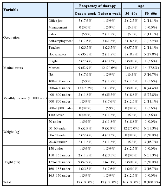
Sample characteristics based on the frequency of back fascia therapy and age (n=34)
2. 족문 측정 결과
실험을 실시하기 전에 족문기에 의한 측정값인 족장Ⅰ, 족장Ⅱ, 족폭, 종골폭, 종골각, 아치각, 모지각, 족선각, 외번각들이 관리 빈도와 연령에 대해서 동질한지를 알아보기 위해 독립표본 t 검정을 실시하였으며 동질한 집단이라는 결과를 얻었다.
1) 족장Ⅰ에 대한 변화 비교
등 근막이완요법이 왼발의 족장Ⅰ에 미치는 영향을 알아보기 위해 기술통계와 반복측정 이원분산분석을 실시하였다. 그 결과를 보면, 왼발의 족장Ⅰ에서 연령, 관리 빈도, 측정 시기에 대한 주 효과와 연령과 관리 빈도의 상호작용효과, 측정 시기와 연령의 상호작용효과, 측정 시기와 관리 빈도의 상호작용효과, 측정 시기 및 연령과 관리 빈도의 상호작용효과에서 통계적인 유의성이 나타나지 않았다(Appendix 1). 오른발의 족장Ⅰ도 효과에 대한 측정 항목에서 통계적 유의성이 확인되지 않았다(Appendix 2).
2) 족장Ⅱ에 대한 변화 비교
등 근막이완요법이 왼발의 족장Ⅱ에 미치는 영향을 알아보기 위해 기술통계와 반복측정 이원분산분석을 실시하였다. 이에 결과를 살펴보면, 왼발의 족장Ⅱ는 연령, 관리 빈도, 측정 시기의 주 효과와 연령과 관리 빈도의 상호작용효과, 측정 시기와 연령의 상호작용효과, 측정 시기와 관리 빈도의 상호작용효과, 측정 시기 및 연령과 관리 빈도의 상호작용효과에서 통계적인 유의성이 나타나지 않았다(Appendix 3).
이에 반해 오른발의 족장Ⅱ에 대한 기술통계를 보면, 전반적으로 30–40대보다 50–60대의 족장Ⅱ의 길이가 길고 시간에 따른 변화폭도 50–60대에서 좀더 컸다(Figure 2). 반복측정 이원분산분석의 결과에서도 측정 시기(F =3.903; p <0.05)에 대해 주 효과가 나타났기에 통계적으로 유의미한 차이를 알아보기 위해 Bonferroni로 조정한 다중비교를 하였다. 실험 전과 실험 1회 후까지는 차이가 보였지만 1회 이후로는 차이가 나타나지 않았다. 이를 통해 실험 1회 이후의 효과가 6회까지 지속되었다는 것을 알 수 있다(Table 2). 반면에 연령, 관리 빈도의 주 효과와 연령과 관리 빈도의 상호작용효과, 측정 시기와 연령의 상호작용효과, 측정 시기와 관리 빈도의 상호작용효과, 측정 시기 및 연령과 관리 빈도의 상호작용효과는 통계적으로 유의미하지 않은 것으로 나타났다(Appendix 4). 이것은 등 근막이완요법의 시행이 연령이나 관리 빈도에 관계없이 족장Ⅱ의 변화에 효과가 있다는 것을 의미하는 것이다.

Descriptive analysis of foot length II of the right foot.
Foot length II of the right foot was greater in women in their 50–60s than in those in their 30–40s. The effects of before vs. after therapy were greater in women in their 50–60s than in those in their 30–40s.
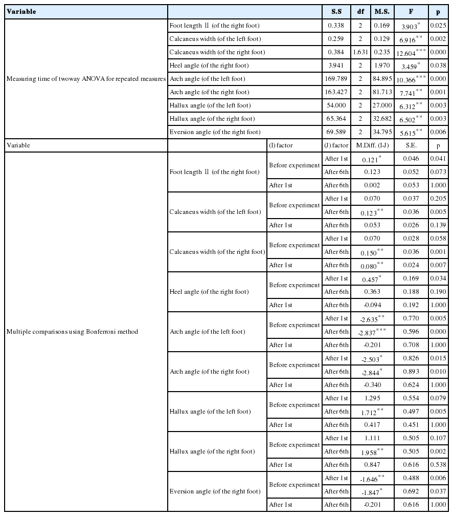
Two–way ANOVA of repeated measures and multiple comparisons using the Bonferroni method on effects of back fascia relaxation therapy
3) 족폭에 대한 변화 비교
등 근막이완요법이 왼발의 족폭에 미치는 영향을 알아보기 위해 기술통계와 반복측정 이원분산분석을 실시하였다. 그 결과는 연령, 관리 빈도, 측정 시기의 주 효과와 연령과 관리 빈도의 상호작용효과, 측정 시기와 연령의 상호작용효과, 측정 시기와 관리 빈도의 상호작용효과, 측정 시기 및 연령과 관리 빈도의 상호작용효과에서 통계적으로 유의미하지 않았다(Appendix 5). 오른발의 족폭에 대한 측정 항목에서도 통계적인 유의성이 나타나지 않았다(Appendix 6).
4) 종골폭에 대한 변화 비교
등 근막이완요법이 왼발의 종골폭에 미치는 영향을 알아보기 위해 기술통계와 반복측정 이원분산분석을 실시하였다. 기술통계를 보면, 30–40대보다 50–60대 변화의 폭이 컸으며, 실험 전부터 6회까지 순차적으로 50–60대의 종골폭이 감소하였다(Figure 3A). 반복측정 이원분산분석을 통해서는 측정 시기(F =6.916; p <0.01)의 주 효과가 나타났다. 측정 시기 간의 통계적 유의미성을 알아보기 위해 Bonferroni로 조정하여 다중비교를 한 결과, 왼발의 종골폭이 실험 1회까지는 효과가 나타나지 않았지만 실험 6회부터 효과가 나타났다(Table 2). 이에 반해 연령, 관리 빈도의 주 효과와 연령과 관리 빈도의 상호작용효과, 측정 시기와 연령의 상호작용효과, 측정 시기와 관리 빈도의 상호작용효과, 측정 시기 및 연령과 관리 빈도의 상호작용효과는 통계적으로 유의미한 차이가 나타나지 않았다(Appendix 7).

Descriptive analysis of the calcaneus width in both feet.
(A) Calcaneus width of the left foot. Effects of therapy on the calcaneus width of the left foot were greater in women in their 50–60s than in those in their 30–40s, and the calcaneus width of the left foot consecutively decreased from before the start of therapy to after the sixth round of therapy. (B) Calcaneus width of the right foot. The calcaneus width of the right foot in women in their 30–40s and 50–60s consecutively decreased from before the start of therapy to after the sixth round of therapy. Moreover, change in the calcaneus width of the right foot was the greatest in women in their 50–60s undergoing therapy once a week.
오른발의 종골폭에 대한 기술통계를 보면, 30–40대와 50–60대 모두 실험 전에서부터 6회까지 종골폭이 순차적으로 감소하였는데 50–60대의 1회군에서 변화의 폭이 가장 크게 나타났다(Figure 3B). 또한, 반복측정 이원분산분석의 결과에서는 측정 시기(F =12.604; p <0.001)의 주 효과가 나타났다. 따라서 측정 시기 간의 통계적 유의미성을 알아보기 위해 Bonferroni로 조정한 다중비교의 결과를 살펴보았다. 실험 전과 실험 1회 간에는 차이가 나타나지 않았지만 실험 전과 실험 6회 사이 그리고 실험 1회와 실험 6회 간에서 유의성 있는 효과가 보였다. 이를 통해 실험에 대한 효과가 실험 1회 이후에 나타났음을 알 수 있다(Table 2). 이에 반해 연령, 관리 빈도의 주 효과와 연령과 관리 빈도의 상호작용효과, 측정 시기와 연령의 상호작용효과, 측정 시기와 관리 빈도의 상호작용효과, 측정 시기 및 연령과 관리 빈도의 상호작용효과에서는 통계적으로 유의미한 차이가 나타나지 않았다(Appendix 8).
이것을 통해 등 근막이완요법의 효과가 종골폭에서는 일정 횟수 후에 나타나며 좌우가 동시에 나타나는 것으로 보아 시행 방법과 발 사이의 관련성이 높은 것으로 짐작된다.
5) 종골각에 대한 변화 비교
등 근막이완요법이 왼발의 종골각에 미치는 영향을 알아보기 위해 기술통계와 반복측정 이원분산분석을 실시하였다. 그 결과, 연령과 관리 빈도, 측정 시기의 주 효과와 연령과 관리 빈도의 상호작용효과, 측정 시기와 연령의 상호작용효과, 측정 시기와 관리 빈도의 상호작용효과, 측정 시기 및 연령과 관리 빈도의 상호작용효과에서 통계적으로 유의미한 차이가 나타나지 않았다(Appendix 9).
이에 반해 오른발의 종골각에서는 효과가 나타났다. 먼저 기술통계를 보면, 30–40대에서는 주1회군이, 50–60대에서는 주2회군이 실험 전에서 실험 6회까지 순차적으로 종골각이 감소하였다(Figure 4). 반복측정 이원분산분석의 결과에서는 측정 시기(F =3.459; p <0.05)에 따른 주 효과가 나타났기에 통계적 유의성을 알아보기 위해 Bonferroni로 조정한 다중비교를 하였다. 오른발의 종골각은 실험 전과 실험 1회 간의 차이가 나타났지만 1회 이후로는 차이가 존재하지 않아 1회의 효과가 6회까지 지속되었음을 알 수 있다(Table 2). 이와 달리, 연령, 관리 빈도의 주 효과와 연령과 관리 빈도의 상호작용효과, 측정 시기와 연령의 상호작용효과, 측정 시기와 관리 빈도의 상호작용효과, 측정 시기 및 연령과 관리 빈도의 상호작용효과는 통계적으로 유의미한 차이가 보이지 않았다(Appendix 10). 이로써 등 근막이완요법의 시행이 연령이나 관리 빈도와 상관없이 종골각의 변화에 효과가 있었음을 알 수 있다.

Descriptive analysis of the heel angle of the right foot.
The heel angle of the right foot in women in their 30–40s undergoing therapy once a week and in those in their 50–60s undergoing therapy twice a week consecutively decreased from before the start of therapy to after the sixth round of therapy.
6) 아치각에 대한 변화 비교
등 근막이완요법이 왼발의 아치각에 미치는 영향을 알아보기 위해 기술통계와 반복측정 이원분산분석을 실시하였다. 기술통계를 살펴보면, 50–60대가 순차적으로 아치각이 증가하였는데 주1회군에서 좀더 변화의 폭이 컸다(Figure 5A). 반복측정 이원분산분석을 통한 결과를 보면, 측정 시기(F =10.366; p <0.001)에서 주 효과가 나타났고 측정 시기에 대한 통계적 유의성을 알아보기 위해 Bonferroni로 조정된 다중비교를 시행하였다. 이에 실험 전에 대해 실험 1회와 실험 6회는 각각 유의성이 확인되었지만 실험 1회에 대해 실험 6회는 차이가 존재하지 않아 실험 1회에서 나타난 효과가 6회까지 지속된 것을 알 수 있다(Table 2). 이에 반해 연령, 관리 빈도의 주 효과와 연령과 관리 빈도의 상호작용효과, 측정 시기와 연령의 상호작용효과, 측정 시기와 관리 빈도의 상호작용효과, 측정 시기 및 연령과 관리 빈도의 상호작용효과에서는 통계적으로 유의미한 차이가 보이지 않았다(Appendix 11).
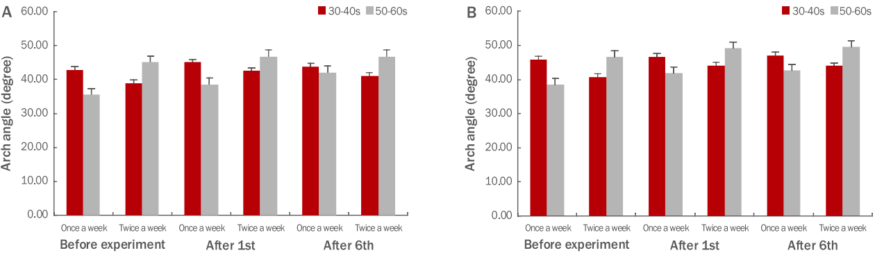
Descriptive analysis of the arch angle in both feet.
(A) Arch angle of the left foot. The arch angle of the left foot in women in their 50–60s consecutively increased from before the start of therapy to after the sixth round of therapy. In particular, effects of therapy on the arch angle of the left foot in women in their 50–60s undergoing therapy once a week were greater than in those in their 50–60s undergoing therapy twice a week. (B) Arch angle of the right foot. The arch angle of the right foot in women in their 30–40s and in those in their 50–60s increased from before the start of therapy to after the sixth round of therapy. Moreover, the change was the greatest in women in their 50–60s undergoing therapy once a week.
오른발의 아치각에 대한 기술통계를 보면, 30–40대와 50–60대에서 실험 전에 비해 아치각이 증가하였는데 50–60대의 주1회군에서 가장 크게 변화하였다(Figure 5B). 반복측정 이원분산분석을 통한 결과를 보면, 측정 시기(F =7.741; p <0.01)에 대한 주 효과가 나타났다. 이에 통계적 유의성을 알아보기 위해 Bonferroni로 조정된 다중비교를 시행한 결과, 실험 전과 실험 1회에서 차이가 나타났다. 그리고 실험 6회에 대해서도 유의성이 존재하였으나 실험 1회와 실험 6회에서는 차이가 존재하지 않아 실험 1회에서 나타난 효과가 6회까지 지속되었음을 알 수 있다(Table 2). 이와 달리 연령, 관리 빈도의 주 효과와 연령과 관리 빈도의 상호작용효과, 측정 시기와 연령의 상호작용효과, 측정 시기와 관리 빈도의 상호작용효과, 측정 시기 및 연령과 관리 빈도의 상호작용효과는 통계적으로 유의미하지 않게 나타났다(Appendix 12).
이 결과를 통해 근막이완요법의 효과가 아치각에서 즉각 나타나고 좌우에서 모두 나타남을 볼 때 등 근막이완요법과 관련성이 높다는 것을 알 수 있다. 실제로 아치각은 발반사구 이론에서 척추와 관련된 위치이며, Song et al. (2010)의 연구에서도 본 연구결과와 동일하게 등 근막관리로 좌우 발의 아치각 값이 커졌다고 보고되었다.
7) 모지각에 대한 변화 비교
등 근막이완요법이 왼발의 모지각에 미치는 영향을 알아보기 위해 기술통계와 반복측정 이원분산분석을 실시하였다. 기술통계를 통해서는 50–60대가 30–40대보다 모지각이 크다는 것과 30–40대의 주1회군과 주2회군은 모두 순차적으로 모지각이 감소한다는 것을 알 수 있었다(Figure 6A). 반복측정 이원분산분석의 결과에서는 측정 시기(F =6.312; p <0.01)에서 주 효과가 나타났기에 통계적 유의성을 알아보기 위해 Bonferroni로 조정된 다중비교를 시행하였다. 실험 전과 실험 6회 사이에 차이가 존재하여 실험의 효과가 나타났음을 알 수 있었다(Table 2). 이에 반해 연령, 관리 빈도의 주 효과와 연령과 관리 빈도의 상호작용효과, 측정 시기와 연령의 상호작용효과, 측정 시기와 관리 빈도의 상호작용효과, 측정 시기 및 연령과 관리 빈도의 상호작용효과는 통계적으로 유의미한 결과가 보이지 않았다(Appendix 13).
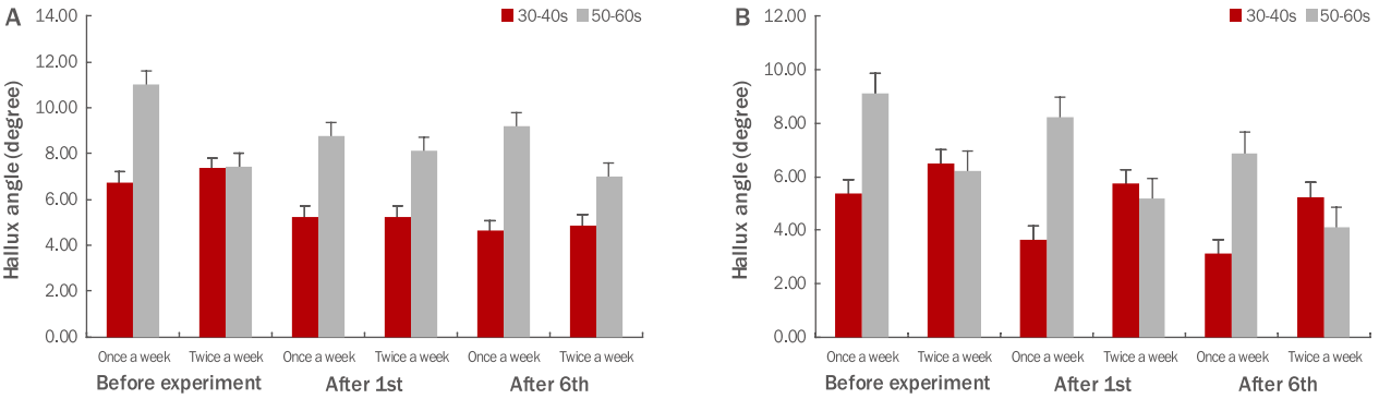
Descriptive analysis of the hallux angle in both feet.
(A) Hallux angle of the left foot. The hallux angle of the left foot in women in their 50–60s was larger than that in those in their 30–40s. The hallux angle of the left foot in women in their 30–40s consecutively decreased from before the start of therapy to after the sixth round of therapy. (B) Hallux angle of the right foot. The hallux angle of the right foot in women in their 30–40s and that in those in their 50–60s consecutively decreased from before the start of therapy to after the sixth round of therapy. Moreover, the change was the greatest in women in their 30–40s undergoing therapy once a week.
오른발의 모지각에 대한 기술통계를 살펴보면, 30–40대와 50–60대 모두 실험 전에서부터 6회 이후까지 순차적으로 감소하였는데 30–40대 주1회군에서 가장 크게 변화하였다(Figure 6B). 반복측정 이원분산분석의 결과에서는 측정 시기(F =6.502; p <0.01)에 따른 주 효과가 나타났다. 이에 Bonferroni로 조정된 다중비교를 통해 통계적 유의성을 살펴본 결과, 실험 전과 실험 6회 간에 차이가 존재하여 실험에 따른 효과가 나타났음을 알 수 있었다(Table 2). 이에 반해 연령, 관리 빈도의 주 효과와 연령과 관리 빈도의 상호작용효과, 측정 시기와 연령의 상호작용효과, 측정 시기와 관리 빈도의 상호작용효과, 측정 시기 및 연령과 관리 빈도의 상호작용효과에서는 통계적 유의성이 보이지 않았다(Appendix 14).
이를 통해 등 근막이완요법이 모지각의 변화에 영향을 준다는 것을 알 수 있었다. 모지각도 아치각과 함께 발반사구에서 척추에 대응하는 위치에 있어 등과 발의 특정 부위가 관련 있음을 확인할 수 있었다. 그리고 Song et al. (2010)의 연구와 같이 본 연구에서도 모지각이 유의하게 감소하였다.
8) 족선각에 대한 변화 비교
등 근막이완요법이 왼발의 족선각에 미치는 영향을 알아보기 위해 기술통계와 반복측정 이원분산분석을 실시하였다. 결과를 보면 연령, 관리 빈도, 측정 시기의 주 효과와 연령과 관리 빈도의 상호작용효과, 측정 시기와 연령의 상호작용효과, 측정 시기와 관리 빈도의 상호작용효과, 측정 시기 및 연령과 관리 빈도의 상호작용효과 모두에서 통계적으로 유의미한 차이가 나타나지 않았다(Appendix 15). 오른발의 족선각에서도 효과에 대한 측정 항목들에서 통계적인 유의성이 보이지 않았다(Appendix 16).
이와 같은 결과는 Song et al. (2010)의 연구와 상반되는 결과로 선행 연구가 10회 관리 횟수였는데 반해 본 연구에서는 6회로 진행되었다는 점에서 비롯한 것으로 짐작된다. 따라서 시행에 따른 효과를 측정하기 위해서는 시행 횟수가 중요함을 알 수 있었다.
9) 외번각에 대한 변화 비교
등 근막이완요법이 왼발의 외번각에 미치는 영향을 알아보기 위해 기술통계와 반복측정 이원분산분석을 실시하였다. 결과는 연령, 관리 빈도, 측정 시기의 주 효과와 연령과 관리 빈도의 상호작용효과, 측정 시기와 연령의 상호작용효과, 측정 시기와 관리 빈도의 상호작용효과, 측정 시기 및 연령과 관리 빈도의 상호작용효과가 통계적으로 유의미하지 않은 것으로 나타났다(Appendix 17).
이와 달리 오른발의 외번각은 효과가 나타났다. 먼저 기술통계를 살펴보면, 30–40대와 50–60대 모두 실험 전과 실험 후 간에 외번각이 증가하였다(Figure 7). 반복측정 이원분산분석의 결과에서는 측정 시기(F =5.615; p <0.01)의 주 효과가 나타났다. 통계적 유의성을 알아보기 위해 Bonferroni로 조정된 다중비교를 시행하였는데, 실험 전과 실험 1회 사이와 실험 전과 6회 간의 차이는 존재하지만 실험 1회와 실험 6회 사이의 유의성은 나타나지 않았다. 이것으로 실험 1회에서 나타난 효과가 6회까지 지속되었다는 것을 알 수 있다(Table 2). 이에 반해 연령, 관리 빈도의 주 효과와 연령과 관리 빈도의 상호작용효과, 측정 시기와 연령의 상호작용효과, 측정 시기와 관리 빈도의 상호작용효과, 측정 시기 및 연령과 관리 빈도의 상호작용효과에서는 통계적으로 유의미한 차이가 보이지 않았다(Appendix 18).

Descriptive analysis of the eversion angle of the right foot.
The eversion angle of the right foot in women in their 30–40s and those in their 50–60s increased from before the start of therapy to after the sixth round of therapy.
이와 같은 결과는 Song et al. (2010)의 연구에서 좌우 발에 모두 유의한 변화가 있었던 것과 차이가 있으며 시행된 횟수의 차이에 의한 것으로 추측된다.
Conclusion
본 연구는 등 부위에 근막이완요법을 시행한 후 발 형태의 변화가 있는지를 알아보고자 하였다. 결과를 통해서 등 근막이완요법이 발 형태를 변화시킨다는 것을 알 수 있었다. 족문에서 종골폭 및 아치각과 모지각은 왼발과 오른발 모두에서 측정 시기에 따른 차이가 나타났다. 족장Ⅱ, 종골각, 외번각은 오른발에서만 유의미한 차이가 존재하였는데 왼발에서만 유의미한 차이가 존재하는 결과가 없는 것은 눈여겨볼 만하다. 그리고 족장Ⅰ, 족폭, 족선각에서는 두 발 모두에서 유의미한 차이가 존재하지 않았다.
연구 결과에서 측정 시기에 따른 유의성은 관찰되었지만 관리 빈도와 연령에 따른 유의성은 나타나지 않아 연령과 관리 빈도에 관계없이 시행 자체가 가지고 있는 효과를 알 수 있었다. 이를 통해 성인 여성들은 등 관리를 통해 등뿐만 아니라 발 관리의 효과를 누릴 수 있으며 시간과 경제적 상황에 따라 최적의 관리 계획을 세울 수 있다. 또한 발반사와 근막경선이론에 따른 인체의 유기적 관계를 동시에 확인할 수 있었다.
지금까지 연구를 살펴보면 등 부위의 관리를 통해 족문의 변화를 살펴본 연구도 드물었지만 스웨디쉬나 경락 등의 기법을 시행한 후 족문의 변화를 관찰한 연구도 제대로 이루어지지 않고 있다. 이에 시행 기법에 따른 연구와 성별에 따른 연구의 필요성을 제기하며 향후 등과 발의 유기적 관계에 대한 심도 있는 연구들이 활발히 진행되길 바란다.