아이 쉐도우 Red/Blue Green의 면적비 변화에 따른 뇌인지
Effect of Changes in the Area Ratio of Red/Blue Green Eyeshadow on Cognitive Processing in the Brain
根据眼影红/蓝绿面积比的变化进行大脑识别
Article information
Abstract
목적
아이 쉐도우 red (R)와 blue green (BG)의 면적비 변화에 따른 뇌파분석을 통하여 아이 쉐도우 색상변화에 따른 뇌파반응을 정량적 데이터를 구축하기 위하여 뇌파(EEG)와 시각유발전위(VEPs) 측정을 하였다.
방법
20대 남녀 60명을 대상으로 red (R), blue green (BG) 아이 쉐도우를 이용하여 면적의 변화를 주어 자극물을 제시하여 자발뇌파(EEG)와 시각유발전위(VEPs)를 측정하여 면적변화에 따른 뇌파반응을 분석하였다.
결과
색상 자극에 대한 뇌파반응은 후두엽 부분이 가장 활성되었다. 특히 아이 쉐도우의 면적변화에 따른 뇌파반응 결과는 황금비율(1.61)일 때 심리적으로 가장 이완되었으며, 단색은 BG 아이 쉐도우에서 α파의 활성 반응으로 가장 안정된 반응이 파악되었다. R 아이 쉐도우만을 사용한 자극물은 각성과 주의력이 높은 것으로 뇌파반응이 도출되었다. 색상대비 시 주조색과 포인트색의 비율에 따라 지각반응이 다르게 작용하였으며, 고채도 일 경우 시각유발전위(VEPs) 반응이 빠르게 작용하였다. 또한 뇌파반응 결과와 색상 이미지가 동일하게 도출된 것으로 보아 상관관계가 높다는 것을 증명하였으며, 적당한 색의 면적 비를 활용할 경우 색상 이미지의 효과를 극대화할 수 있음을 증명하였다.
결론
본 연구결과는 컬러의 면적 비를 조절하여 색채계획을 세울 경우 원하는 색상의 효과를 과학적으로 도출할 수 있었다. 아이 쉐도우 면적변화에 따른 도출된 뇌인지반응은 이미지 메이킹 시 정량적 데이터로 활용하여, 뷰티산업 분야에서 다양하게 활용도를 넓일 수 있을 것으로 사료된다.
Trans Abstract
Purpose
To conduct electroencephalography (EEG) and visual evoked potentials (VEPs) measurements to establish quantitative data on brainwave responses to changes in eyeshadow color, specifically the area ratio of red (R) and blue green (BG) eyeshadows.
Methods
A study was conducted on 60 males and females in their 20s using R, BG eyeshadow to determine brainwave responses via spontaneous EEG and VEPs while presenting stimuli with changes in area. The analysis focused on brainwave responses to changes in area.
Results
The brainwave responses to color stimuli demonstrated the maximum activity in the occipital lobe. Specifically, the results of the EEG analysis indicated the highest level of psychological relaxation response at the golden ratio (1.61) of the eyeshadow area. Furthermore, when participants wore BG eyeshadow, their brainwave responses remained stable, with consistent alpha wave activation. The presentation of stimuli using only the R eyeshadow resulted in a significant increase in arousal and attention levels among the participants. Perceptual responses exhibited variations based on the ratio of the main color and point color in the color contrast, and rapid VEPs responses were observed specifically under high saturation conditions. Furthermore, the study revealed a high correlation between the brainwave response results and color images, indicating that an optimal ratio of color areas can maximize the impact of the color image.
Conclusion
The results of this study suggest that by adjusting the area ratio of colors, it is possible to scientifically determine and achieve the desired color effects in color planning. The brain cognitive responses, resulting from variations in eyeshadow area, can serve as valuable quantitative data for image-making purposes, and can be applied to various fields in the beauty industry to expand their usability
Trans Abstract
目的
进行脑电图(EEG)和视觉诱发电位(VEP)测量,以建立脑电波对眼影颜色变化反应的定量数据,特别是红色(R)和蓝绿色(BG)眼影的面积比。
方法
对60名20多岁的男性和女性进行了一项研究,使用 R、BG眼影,通过自发EEG和VEP确定脑电波反应,同时呈现面积变化的刺激。分析的重点是脑电波对区域变化的反应。
结果
对颜色刺激的脑电波反应显示枕叶的最大活动。具体来说,脑电图分析结果表明,眼影区域黄金比例(1.61)处的心理放松反应水平最高。此外,当参与者佩戴 BG 眼影时,他们的脑电波反应保持稳定,α 波激活一致。仅使用 R 眼影呈现刺激会显着提高参与者的唤醒度和注意力水平。感知反应表现出基于颜色对比度中主色和点色的比例的变化,并且特别是在高饱和度条件下观察到快速的 VEP 反应。此外,研究还揭示了脑电波响应结果与彩色图像之间的高度相关性,表明最佳的彩色区域比例可以最大限度地发挥彩色图像的影响。
结论
本研究结果表明,在色彩规划中,通过调整色彩的面积比例,可以科学确定并达到理想的色彩效果。由眼影区域变化产生的大脑认知反应可以作为图像制作目的的有价值的定量数据,并且可以应用于美容行业的各个领域以扩展其可用性。
Introduction
색은 보고 느끼는 감성적 표현이 개개인에 따라 다양하게 나타나며, 색 자극에 따라 사람의 기분이나 생활, 분위기, 환경 등에 영향을 준다. 특히 색에 대한 특정한 경험과 기억은 사람의 감성을 움직이는 큰 역할을 한다(Birren, 1984). 색 이미지는 한가지의 색 만을 보고 느낄 때와 달리 여러가지 색이 조화되면 원래색이 가지고 있는 이미지와 다르게 느끼게 된다. 색을 보고 느끼는 감성반응은 개인의 기호나 직관에 의하여 결정되는 것이 아니라 색채이론과 기능, 심리학, 생리학, 미학, 색채 반응 등과 연관되어 작용된다. 배색된 색은 색의 속성(색상, 명도, 채도)에 의하여 배색조건이 바뀌게 되어 원래 색 이미지와는 다르게 지각된다. 색상은 동일배색이나 유사배색, 보색배색 등에서 조화가 잘 되었을 경우 미적으로 우수하고 강한 느낌을 준다(Kim & Jeong, 2008). 이와 같은 배색은 주변색과 어우러져 도출된 감성연구보다 각각의 단색의 특성 또는 색 이미지만을 평가하는 경우가 다수였으며(Ainsworth et al., 1993; Goodfellow & Smith, 1973; Green et al., 1982), 특히 단색 red에 대한 색 이미지를 단편적으로 느껴지는 이미지대로 정성적 평가를 한 연구들이 다수였다(Isaacs, 1980; Kwallek & Lewis, 1990; Shick, 1975). 색을 보고 느끼는 감성 반응은 강렬, 열정적, 편안함, 시원함 등의 단편적 이미지 감성을 느낄 수 있다(James & Domingos, 1953; Mehta & Zhu, 2009; Nakashian, 1964; Sinclair et al., 1998; Elliot, 2015). 색은 생리학적 효과를 주며 사람의 행동에 긍정적 또는 부정적인 영향을 주고(Dittrichová et al., 1989; Ansorge et al., 2009), 신체의 체온변화(Noguchi & Sakaguchi, 1999), 수면을 유도(Kozaki et al., 2005), 집중력 향상에 도움을 주며(Ecker et al., 2007), 시각자극에 영향을 줌으로 사람의 정서에 영향을 주는 것으로 파악되었다(Paulus et al., 1984). 그림의 색으로 인하여 기억력의 증감현상(Cycowicz et al., 2008), 컴퓨터의 바탕색에 따른 집중력 향상(Boucsein et al., 2001; TaKano et al., 2009)에 영향을 주는 정서적 반응에 대한 연구도 있다. 또한 색광자극이 사람의 생체반응(뇌파측정, 심전도)을 통하여 생리적으로 영향을 주는 정량화 된 연구가 진행되었다(KÜller, 1986; Kobayashi & Sato, 1992; Küller, 2002; Yeh et al., 2013). 그 외 인테리어 색상에 따라 감성 반응이 다양하게 나타나는 결과(Küller et al., 2009)를 통하여 색이 실제 생활에 적용되었을 경우 심리적 변화를 수반함을 증명하였다. 그러나 이러한 기존 연구는 컬러를 이용하여 사람의 심리 · 생리적 반응을 나타낸 연구로, 일부 한정된 컬러의 자극물을 제시하여 나타낸 결과이므로 색상이 어울려 느끼는 반응들과 면적 비의 변화에 따른 자극결과는 알 수 없었다. 아이 쉐도우 색상을 보고 느끼는 즉각적인 반응을 정량적 결과로 도출하기 위하여 시각유발전위 검사는 시각회로의 신경생리적 기능을 평가할 수 있으므로 시각 기능의 연구에 있어 매우 중요한 도구이다(Aminoff & Goodin, 1994). 따라서 본 연구는 아이 쉐도우 색상을 포인트색과 바탕색의 면적비를 변화를 주어 자극물로 제시하여 느껴지는 뇌인지 반응을 자발뇌파(EEG)와 시각유발전위(VEPs)를 통하여 분석하였다. 아이 쉐도우 red (R)와 blue green (BG)의 면적비에 따라 생리반응을 정량화하기 위하여 뇌파측정을 하였다. 아이 쉐도우 면적변화에 따른 뇌파반응은 뷰티산업 뿐만 아니라 다양한 분야에 정량화된 결과를 활용도를 높이고자 시도하고자 한다.
Materials and Methods
1. 실험대상 및 자극물
아이 쉐도우는 R (5R vivid, 5/14)와 보색 관계인 BG (5BG vivid, 5/6)를 사용하여 일러스트북 이미지를 활용하여 시각자극물은 제작하였다(Kim et al., 2018). Red (R)와 blue green (BG)을 바탕색과 포인트색으로 사용하여 각각의 바탕색과 포인트색의 면적 비(A 바탕색/A포인색)를 1.42, 1.61, 5, 8 그리고 A포인색/Atot를 0.41, 0.38, 0.17, 0.11, 0으로 변화시켰다(Table 1). 아이 쉐도우 제품은 R은 색연필과 에리조 제품의 레드 #1 컬러를 사용하였고, BG은 색연필과 에리조 제품의 에메랄드 그린 아이즈 #86 컬러를 사용하여 시각자극물을 제작하였다(Table 1). 눈과 눈썹에만 컬러링하였고 완성된 자극물들을 스캔하였다. 스캔 된 자극물은 피험자들에게 모니터로 제시하였다. 이들 시각자극물에 대한 뇌파 반응은 서울 경기도 소재의 20대 남녀 대학생, 남성 30명(평균25.2±0.8세), 여성 30명(평균23.2±0.7세)을 대상으로 실험을 진행하였다. 실험대상자중 바탕 뇌파가 불안정한 남성 3명, 여성 2명은 배제하였고, 뇌파의 좌 · 우측의 대칭된 정상 뇌파 패턴을 가진 최종 55명을 선정하였다. 피시험자들은 beck depression inventory (BDI) 측정을 통하여 신경학 또는 정신질환 병력이나 색맹이 아닌 인지장애 없는 대상만을 선발하였다. 본 연구는 헬싱키윤리기준과 임상시험기준을 준수했으며, 보건복지부 지정 공용기관 인 생명윤리위원회의 승인을 받았다(승인 번호: P01201807-11-002). 측정공간은 실내 조도와 온도를 각각 150 lux와 23-24 oC로 일정하게 유지시키고 항온/항습 공간에서 선정된 시각자극물을 이용하여 피시험자들의 뇌파 반응을 측정하였다.

Color stimuli used for the complementary contrast of red and blue green eyeshadows
2. 실험 방법
1) EEG 측정
뇌파 생리신호(EEG)는 QEEG 32system (Laxtha Inc., Korea)을 사용하여 측정하였고 뇌파 측정부위는 국제 10-20 전극배치법의 기준에 따라 19부위(Fp1, Fp2, F3, F4, F7, F8, Fz, T3, T4, C3, C4, Cz, P3, P4, Pz, T5, T6, O1, O2)에 부착하여 단극 유도법으로 측정했다. 준비된 시각자극물은 모니터를 통하여 제시하였다. 바탕뇌파는 눈을 감은 상태에서 자극 없는 편안한 상태에서 30 s 동안 뇌파를 측정하였다. 안구 움직임이나 안면근육 수축 등과 같은 몸의 움직임이 없음에도 불구하고 바탕뇌파의 변동이 심한 피시험자는 배제시켰고 정상적인 바탕뇌파가 측정된 피험자를 대상으로 하였다. 뇌파측정 전 피시험자가 안정상태를 유지시켜 눈을 감은 체 10-20 s 동안 명상을 취한 후, 눈을 뜨고 색상 자극물이 있는 모니터를 15 s 동안 응시하도록 한 상태에서 뇌파를 수집하였다. 모니터와 피험자와의 거리는 50 cm정도의 거리를 두었고, 피시험자에게 시각 자극물의 노출순서는 무작위로 하였다.
2) VEPs 측정
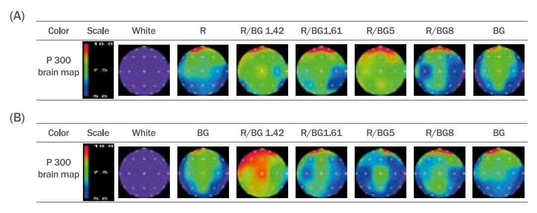
뇌파 생리신호(EEG)는 QEEG 32 system (Laxtha Inc., Korea)을 사용하여 측정하였고 뇌파 측정부위는 인지유발전위(cognitive evoked potential, P300)의 활성전극은 국제 10-20 system 중 Fz, Cz, Pz로 정하였고 보조 전극은 양쪽 귓 볼(A1, A2)에 위치하였다. Brain map은 국제 10-20 전극배치법의 기준에 따라 19부위(Fp1, Fp2, F3, F4, F7, F8, Fz, T3, T4, C3, C4, Cz, P3, P4, Pz, T5, T6, O1, O2)에 부착하여 단극 유도법으로 측정하여 도시하였다(Figure 4, Figure 5).

VEP responses shown according to changes in the area of the red background color (R) and focal color (BG).
Stimuli were presented with six eye-shadow colors, including no eyeshadow, single-color eyeshadows on R and BG backgrounds, and two-color eye-shadows with area changes of 1.42, 1.61, and 8. P300 waveforms were obtained for each eye-shadow color. (A) No eyeshadow; (B) Red; (C) R8BG; (D) R5BG; (E) R1.61BG; (F) R1.42BG; (G) BG.

VEPs response to variations in the area of the background color (BG) and the focal color red.
Stimuli were presented with variations in the area of single-color BG and red eye-shadows as well as in the form of two different colors at area changes of 1.42, 1.61, and 8. A no eye-shadow stimulus was also presented. P300 waveforms were obtained for each eye-shadow color. (A) No eyeshadow; (B) BG; (C) BG8R; (D) BG5R; (E) BG1.61R; (F) BG 1.42R; (G) Red.
3. 데이터 분석
1) EEG 분석
수집된 뇌파 데이터는 TeleScanTM (Ver.2.99) 프로그램을 이용하여 분석하였다. 전체 0-50 Hz의 주파수 영역 중에 눈 굴림이나 안면 근육의 움직임에 따라서 영향을 많이 받는 델타파(0-4 Hz) 제거 후 Fast Fourier transform (FFT)의 알고리즘을 이용하여 나머지 주파수 대역(4-50 Hz)의 파워 스펙트럼을 분석하였다. 자극이 없는 상태의 바탕 뇌파(RPVs) 대비 아이 쉐도우 색상변화에 의한 상대 뇌파 파워 차이의 비율인 뇌파변동계수(coefficient of variation for the brainwave, CVB)로 분석하였다. 각 자극에 의한 SEF50 (spectral edge frequency 50%)과 SEF90 (spectral edge frequency 90%) 시각 자극에 의한 spectral edge frequency (SEP50과 SEP90)의 변화는 바탕 뇌파의 평균값 대비 편차로 평가하였다(Kim, 2018; Kim, 2022).
전체 주파수 대역의 theta (4-8 Hz), alpha (8-13 Hz), beta (13-30 Hz), gamma (30-50 Hz)을 분석하였고, spectral edge frequency (SEF)인 SEF50와 SEF90을 분석하였다(Kim, 2018). 시각 자극이 없는 바탕 뇌파의 상대 파워값 대비 시각 자극에 따른 상대 파워값(relative power values, RPV)과 바탕뇌파의 상대 뇌파 파워값의 차이의 비율인 뇌파변동계수(CVB)를 분석하였다.
CVB(%)=(xi-Xi)/Xi×100
여기에서 xi는 자극물에 대한 i-주파수 대역의 상대 뇌파값, Xi는 바탕 뇌파의 i-주파수 대역의 상대 뇌파값이다. 시각 자극에 의한 spectral edge frequency (SEP50과 SEP90)의 변화는 바탕 뇌파의 평균값 대비 편차로 평가하였다.
∆SEF (50 or 90)=SEFi-SEFback
시각 자극에 의한 각종 뇌파지표들의 변화값은 시각자극 시 뇌파지표값과 바탕뇌파 뇌파지표 값의 차이로 구하였다. 여기에서 SEFi는 색상 자극에 의한 SEF이고, SEFback은 바탕 뇌파의 SEF이다.
2) VEPs과 통계분석
색상자극에 따른 유발뇌파는 자극제시 후 250-700 ms 사이에서 출현하는 피크(positive, 상향) P300을 분석하여 내적인 요소에 해당하는 인지반응을 분석하였다. 뇌파와 감성반응에 대한 통계분석은 IBM SPSS24을 사용하여 반복측정 분산분석(Repeated Measure ANOVA)을 실시하였고, 95% 유의수준에서 Tukey test를 통해 사후 검증을 하였다.
Results and Discussion
1. 아이 쉐도우 컬러자극에 따른 EEG
1) CVBs와 SEF
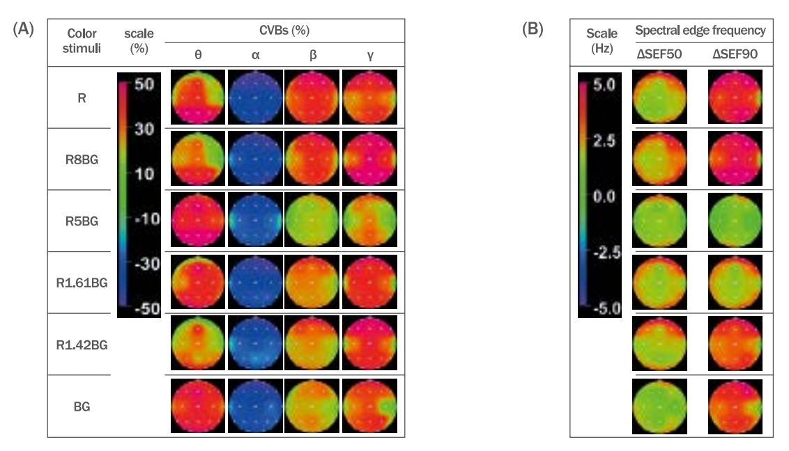
바탕색 R에 포인트색 BG을 단색과 4가지의 면적변화에 의한 자극물을 제시하여 뇌파결과를 Figure 3에 도시하였다. R와 BG의 아이 쉐도우 색상자극에 따른 상대뇌파의 변동계수는 α파를 제외한 나머지 주파수대역에서 바탕 뇌파값 보다 모두 증가하는 추세였다. θ파는 R1.61BG의 자극 시 48.34±10.75%로 가장 많이 증가하였으며 R8BG의 자극 시 가장 작게 증가하였다. α파는 R1.61BG의 자극 시 30.38±8.63%로 가장 작게 감소하였다. 특히 전전두엽은 비활성되었으나 모든 색상자극시 측두엽은 활성되었다. 이완과 안정을 나타내는 α파는 R (-41.61±6.15%)색상의 자극 시 가장 크게 감소하였다. β파는 R의 자극 시 38.19±9.68% 가장 많이 증가하였고, R1.61BG의 자극 시 15.60±7.68%로 가장 비활성 되었다. γ파는 R1.42BG의 자극시 47.95±15.10%로 가장 활성 되었다. 특히 γ파는 색상자극시 전두엽 부위가(50.55%) 활성되었으나 측두엽(T3, T4)은 18.35%로 다른 부위에 비하여 비활성 되었다(Figure 1a).
P300 response according to the area changes of red and BG eye-shadow.
Stimuli with area changes of 1.42, 1.61, and 8 for the solid colors of red and BG eye-shadow, and two colors were presented, and the resulting P300 responses were observed. (A) Results of P300 response to the area changes of BG on the red eye-shadow background. (B) Results of P300 response to the area changes of red on the BG eye-shadow background.
EEG responses to changes in the area of BG and red eye-shadows.
Six different eye-shadow colors were used with variations in their area, and brain wave responses were collected. The stimuli comprised single-color BG and red eye-shadows, as well as variations in their area by a factor of 1.42, 1.61, and 8. The EEG responses to these stimuli were recorded. (A) CVBs results for θ, α, β, and γ waves with changes in the area of red eye-shadow on a BG background. (B) SEF50 and SEF90 results with changes in the area of BG eye-shadow on a red background. R, red color; R8BG, the area of bluegreen compared to the red background color is 8; R5BG, the area of Bluegreen compared to the red background color is 5; R1.61BG, the area of bluegreen compared to the red background color is 1.61; R1.42BG, the area of bluegreen compared to the red background color is 1.42; BG, bluegreen color.
뇌의 활성도 지표인 SEF50과 SEF90는 바탕뇌파 대비 R1.61BG을 자극물로 제시했을 때 △SEF50 (1.62±0.83 Hz), △SEF90 (1.08±0.86 Hz)로 증가 폭이 가장 작았으나 R1.42BG의 자극 시 △SEF50 (3.02±1.05 Hz), △SEF90 (4.74±1.39 Hz)로 가장 증가현상이 두드러졌다(Figure 1b). 단색인 R의 자극 시에도 △SEF50 (2.44±1.07 Hz), △SEF90 (4.42±1.29 Hz)의 증가현상이 두드러졌다. 뇌파지표들의 CVBs값과 △SEF50, △SEF90 모두 통계적으로 유의하였다[θ파, F (6, 437)=97.605, p<0.000; α파, F (6, 437)=472.559, p<0.000; β파, F (6, 437))=204.629, p<0.000; γ파, F (6, 437)=111.767, p<0.000; △SEF50, F (6, 437)=81.796, p<0.000; △SEF90, F (6, 437)=173.260, p<0.000].
바탕색 BG에 포인트색 R를 단색과 4가지의 면적변화에 의한 자극물을 제시하여 뇌파결과를 Figure 2에 도시하였다. BG의 자극 시 θ파는 41.88±8.77%로 가장 활성되었고, 19부위중 전두엽(43.24%)에서 활성반응이 두드러졌다. α파의 CVB는 BG1.61R의 자극 시 33.42±7.38%로 가장 감소폭이 작았고, R 자극 시 감소폭이 컸다. 주로 α파의 CVBs의 반응은 전전두엽에서는 비활성되었으나 측두엽에서는 활성되었다. β파의 CVB는 R자극 시 38.19±9.68%로 가장 활성정도가 두드러졌으며, γ파의 CVB는 BG5R 자극 시 45.74±14.03%로 가장 많이 증가하였다. 특히 전두엽에서 활성반응이 두드러졌다(Figure 2a).

EEG responses to changes in the area of BG and red eye-shadows.
Brain waves were extracted using six different eye-shadow colors with variations in their areas. Stimuli were presented using single colors of BG and red eye-shadows, as well as two colors with area variations of 1.42, 1.61, and 8. The EEG responses to these stimuli were examined. (A) CVBs results for θ, α, β, and γ waves in response to changes in the area of red eye-shadow on a BG background. (B) SEF50 and SEF90 results in response to changes in the area of BG on a red eye-shadow background.
파워값의 중위수인 SEF50과 SEF90의 바탕뇌파 대비 색상자극에 의한 변화량 △SEF50은 BG의 자극 시 가장 작게(14.32±1.60 Hz) 증가한 반면 BG5R이 가장 크게(15.62±1.93 Hz) 증가하였다. R아이 쉐도우 자극 시 스트레스와 각성과 관련 있는 △SEF90가 가장 두드러지게 증가하였고 특히 측두엽을 제외한 전영역에서 활성되었다 (Figure 2b). 뇌파지표들의 CVBs값과 △SEF50, △SEF90 모두 통계적으로 유의하였다[θ파, F (6, 437)=97.605, p<0.000; α파, F (6, 437)=56.955, p<0.000; β파, F (6, 437))=19.391, p<0.000; γ파, F (6, 437)=5.549, p<0.000; △SEF50, F (6, 437)=5.663, p<0.000; △SEF90, F (6, 437)=8.727, p<0.000].
2) 시각유발전위(VEPs)
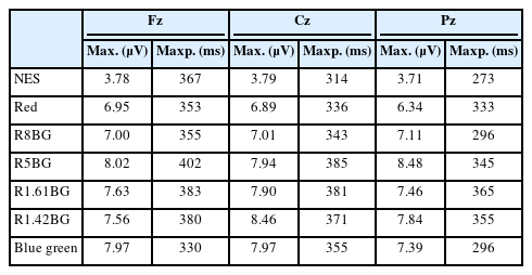
바탕색에 포인트색을 면적 변화한 자극을 통하여 입력된 객체에 대하여 해당 개체를 인식하기까지 걸리는 시간 영역에서의 특징으로 자극 입력 후 약 300-700ms에서 강한 양의 방향 전압(μV)이 발생하는 P300을 Table 2, Table 3와 Figure 3, Figure 4, Figure 5에 도시하였다.
P300 response to color stimuli of contrasting BG color on R eyeshadow background

P300 response to color stimuli of contrasting R color on BG eyeshadow background
다른 색상 자극보다 no eyeshadow (NES)를 볼 때 Cz에서 3.794 μV로 피크값이 가장 작게 출현하였으나, 단색인 R보다 BG의 자극 시 P300의 반응이 Cz의 부위에서 7.967 μV로 높았다. 그러나 면적 비율로 나누어 자극물을 보여주었을 때는 단색의 자극물을 제시했을 때의 반응보다 더 컸다(Table 2, Table 3).
Red가 바탕색이고 포인트색이 BG인 경우 R5BG의 자극 시 8.48 μV로 높게 반응하였다. 그러나 BG이 바탕색이고 red가 대비색일 경우 BG1.42R일 때 9.05 μV로 반응하였다. 색상 자극 시 대상회(cingulate gyrus)부위(Fz, Cz, Pz) 중 Cz에서 가장 크게 증폭되었다(Figure 3). 면적변화 된 자극물을 제시할 경우 바탕색 대비한 포인트색상의 면적이 1.42일 때 가장 인지반응이 최대로 출현하였다(Figure 4). 단색의 경우 시각적 자극이 적은 BG을 자극물로 하였을 경우 빠르게 반응하였다. 바탕색이 R의 경우보다 BG인 경우가 인지반응이 크게 작용하였다(Figure 5). 색상자극 시 인지 반응(P300)은 전전두엽이 두드러지게 활성되었다(Figure 3). P300의 뇌파값은 모든 색상에서 통계적으로 유의하였다[no eyeshadow, F (5, 162)=88.193, p<0.000; R1.42BG, F (5, 162)=39.145, p<0.000; BG1.42R, F (5, 162)=55.736, p<0.000; R5BG, F (5, 162)=39.145, p<0.000; BG5R, F (5, 162)=51.554, p<0.000; R8BG, F (5, 162)=80.700, p<0.000; BG8R, F (5, 162)=35.997, p<0.000; R1.61BG, F (5, 162)=48.436, p<0.000; BG1.61R, F (5, 162)=48.436, p<0.000].
Discussion
R와 BG의 아이 쉐도우 색상을 대비색과 포인트색으로 각각 면적 변화를 주어 뇌파반응을 측정한 결과 Figure 1, Figure 2, Figure 3, Figure 4, Figure 5, Table 2 그리고 Table 3에 도시하였다. Red 색상은 가시광선에서 가장 느린 진동파를 갖고 있으므로 어떤 색보다 가장 빠르고 즉각적으로 감성을 자극하면서 시각적으로도 가장 강하게 영향을 미친다(Shin, 2009; Schmüser et al., 2014). 그러나 R색상과 달리 BG의 색 이미지는 시각적으로 안정되며 편안한 감성을 가지는 색상이다(Shin, 2009). 이 두가지 아이 쉐도우 색상을 자극물로 VEPs측정시 기억탐색과 인지과정을 나타내는 P300의 결과는 R보다 BG의 반응이 더 빠르게 나타났으며, 두 색상을 면적비로 나누어 제시한 색상자극(R5BG, R1.42BG, BG1.42R)은 단색에 비하여 P300이 빠르게 나타났다. 이에 반하여 NES 자극에서는 가장 인지반응이 늦었다(Table 2, Table 3). 이는 고채도와 고명도의 아이 쉐도우 색상인 R가 눈에 자극이 작은 NES의 자극보다 주의력, 기억력, 인지과정이 빠르다는 것으로 유추할 수 있었다. 어린이 공부방의 인테리어 할 경우 산만한 아이들의 정서적 안정을 위하여 한색계통인 blue나 green계열로 디자인한다. 이는 색채가 인체와 감성이 밀접하게 연관되어 있으므로 구현하는 색채에 따라 사람의 감성에 효과적이기 때문이다(Birren, 1973). 이러한 이론과 연구는 BG의 아이 쉐도우 색상 자극에 의한 인지반응인 P300이 이 높게 증폭된 결과를 지지하였다(Table 2, Table 3). Mehta & Zhu (2009)의 의하면 red의 자극 시 기억력을 높이는 효과적이라고 하였고, blue는 창의력과 사람의 감성을 자극하여 작업환경의 증진효과를 줄 수 있다고 하였다. 또 다른 연구에서 purple wheels과 grayscale wheels를 자극물로 하여 지각 테스크(perceptual task)를 실시한 결과 채도가 없는 grayscale wheels보다 color (purple) wheels을 본 반응이 더 빠르게 나타났다(Simmons et al., 2007). 이 현상은 본 연구 결과와 동일하게 무채색(white, black, gray)보다 유채색 아이 쉐도우에서 시각 인지반응이 빨랐다(Figure 4, Figure 5). 본 연구에서는 채도가 높은 red보다 채도가 낮은 BG의 자극에서 P300 반응이 크게 나타난 결과와는 다르게 도출되었다. 체크무늬로 배색된 색상(red와 green)인 경우는 인지가 늦었으나, 본 연구에서는 바탕색 대비 배색의 면적 비가 1.42인 경우는 인지반응이 높다고 유추할 수 있었다(Table 2, Table 3). 그 외 연구에서 white, red and blue light를 flash자극을 준 결과 white는 본 연구 결과와 같이 인지 반응이 작았으며 red와 blue는 거의 비슷하게 도출되었다(Subramanian et al., 2012).
시각유발 반응결과와는 달리 자발뇌파의 반응결과는 BG색상자극보다 red색상의 자극 시 β파와 γ파의 증가폭과 SEF50, SEF90의 반응이 크게 나타났다(Figure 1, Figure 2). 이 결과는 채도가 높은 색상에 대한 뇌의 각성과 활성 정도가 크게 나타난 것으로 파악된다. 즉 눈에 자극적인 R색상으로 인하여 각성반응이 크게 유도함을 파악할 수 있었다. BG바탕색 대비 R아이 쉐도우배색 면적이 5일 때 β파, γ파, SEF50, SEF90의 반응은 뇌의 전 영역에서 활성되었고, R바탕색 대비 BG배색 면적이 1.42일 때 β파, γ파, SEF50, SEF90의 반응도 전 영역에서 증가하였다(Figure 1). 바탕색이 BG의 경우보다 R일 때 시각유발반응과 인지반응이 크게 활성 되었다(Table 2, Table 3). 이러한 결과는 바탕색과 대비색의 면적비율에 따라 색에 대한 인지 정도가 다르다는 것을 유추할 수 있었고, 단색보다는 두 색의 면적변화 줄 경우 지각반응이 각기 다르게 도출할 수 있었다. 본 연구 결과에서 채도가 높은 R 아이 쉐도우 자극 시 BG보다 β파와 γ파, SEF50과 SEF90 반응이 높은 결과를 통하여 채도가 높을수록 시각적 자극을 유도하나, P300의 인지반응은 늦다는 것을 유추할 수 있다. 이는 아이 쉐도우 색상의 면적 비에 따라서 다양한 효과를 유도할 수 있다는 것을 입증하였다.
BG의 색상 이미지는 안정, 편안, 균형, 건강, 젊음 등의 색 이미지로 보는 사람들에게 자극이 없이 편안한 이미지이다(Birren, 1984; Shin, 2009). BG 아이 쉐도우 색상 자극물로 하여 뇌파를 측정한 결과 안락감과 편안함의 뇌파지표인 α파가 가장 높게 증가한 것을 통하여 green계열의 색상이 편안함과 안정감을 준다는 색상 이미지와 일치했다. Kim & Ryu (2011)에 의하면 색상 이미지는 뇌파반응 결과와 밀접한 상관관계가 있다. 본 연구결과에서도 역시 색상 이미지와 뇌파반응과 동일한 경향으로 출현한 결과는 서로 상관관계가 높다고 단정지을 수 있었다.
배색(color scheme)은 목적과 기능에 적합한 효과를 얻기 위하여 여러 색을 계획적으로 배치하는 것으로, 개개의 색채에는 고유성이 강하나 실제로 색채의 독자성에 지각되는 것 보다 인접한 색 이미지에 의하여 좌우되는 경우가 많다(Kuehni, 2003). 그리고 색은 바탕색 또는 대비되는 색은 면적비에 따라 명도와 채도가 다르게 인지되었다(Moon, 2010). 본 연구에서 색의 성질을 이용하여 아이 쉐도우의 바탕색과 포인트색의 면적비를 변화시켜 뇌파 측정 결과 다른 면적 비보다 바탕색대비 포인트색 면적이 1.61일 경우 즉, 전체 색상의 면적 비가 황금비율 일 때 뇌파반응이 이완 정도가 가장 크게 활성되었다. 황금비의 색상자극일때 알파파가 활성 된 결과가 도출되었다. 이 결과는 황금비율이 안정되고 편안하게 지각되는 이론(Andreassi, 2007)을 뒷받침하는 뇌파결과였다(Figure 1, Figure 2). 이러한 이론을 바탕으로 황금비율(황금분할)은 과거부터 현재에 이르기까지 예술 분야, 특히 건축, 미술 등에 즐겨 활용되고 있다. 본 연구에서 시각 자극물에 의한 뇌의 반응이 면적비가 1.61일 때 안정되고 이완된다고 결과가 도출되었으며 우리가 색상을 보고 느끼는 생리학적 반응과 건축, 미술 이론인 황금비율에서 안정감을 느낀다는 이론과 일치하였다. 색상은 대비하는 면적 크기에 따라 다르게 인지되지만, 본 연구에서는 바탕색과 대비색의 위치를 바꾸어도 황금비율에서 안정감이 드는 현상은 동일하였다. 즉 바탕색 대비 대조색의 면적비율이 변화에 따라 상대 뇌파의 비율(relative band power)은 일정한 패턴을 나타냈으며 이 결과는 바탕색과 대조색과의 대비가 크게 느껴졌기 때문이라고 사료된다.
면적에 따른 뇌파반응의 주의, 집중도는 배색된 색상보다 단색일 경우 더 높게 출현했다. 일상 생활에서도 주의를 끌거나 집중시키기 위하여 vivid tone의 red나 yellow red를 많이 사용한다. PCCS tone 분류에 의한 12 tone 중 vivid tone은 가장 주의를 끄는 tone이고 많은 색상 중 red가 가장 주의(awareness)를 끄는 색상이다(Moon, 2010; Birren, 1984). 본 연구에서도 R 아이 쉐도우 자극에서 감마파와 베타파의 활성정도가 높은 것으로 보아 붉은색상의 아이 쉐도우가 자극적이어서 각성도를 높이는 것으로 유추할 수 있었다.
컬러 white, black, gray, red, green, blue, yellow, purple 자극물로 하여 측정한 결과 PO8, Fz, T7, P5에서 반응이 활발했으며(Becker et al., 2009), 색광인 blue, green, red를 자극물로 하여 뇌파를 측정한 결과 Pz에서 가장 높게 반응하였다(Yoto et al., 2007). 뇌의 반응 영역에 관한 또 다른 연구는 occipital lobe에서 볼 수 있는 시각영역이라고 보고되어(Glazov, 2007) 본 연구결과의 색상자극시 후두엽에서 활성반응이 활발하다는 결과를 지지하였다(Figure 3). 픽토그램의 가독성을 살펴본 결과 color combination에 따른 노출시간에 따라 P100과 P300의 반응이 크게 반응하였다. 이 결과는 단색보다 색을 대비시킨 경우 인지 반응이 큰 것을 의미하였다(Yeh et al., 2013). 본 연구 결과에서도 역시 색의 대비에 따른 반응결과가 두드러졌다.
본 연구에서 아이쉐도우 색을 본 뇌의 반응은 색채 이론과 비슷한 결과로 출현하여 기존의 이론을 신뢰할 수 있는 기초를 마련할 수 있었으며, 색채 이미지 이론을 바탕으로 메이크업 색채 계획을 세울 경우 과학적 근거로 제시할 수 있을 것으로 사료된다.
Conclusion
본 연구는 red와 bluegreen의 아이 쉐도우를 면적 변화시켜 자극물로 제시한 뇌파측정결과는 다음과 같았다.
아이 쉐도우 자극에 의한 반응은 뇌의 각 부위 중 후두엽 부분이 가장 활성되었다. 5가지 면적변화(1.42, 1.61, 5, 8)에 따른 뇌파반응 결과는 황금비율(1.61)의 면적일 때 뇌파반응이 가장 이완되었다. 단색의 BG의 아이 쉐도우 자극시 α파가 활성되어 안정된 색상반응으로 도출되었다. 각성과 주의력은 면적대비 된 색보다 단색의 자극에서 반응이 높았고, 그 중 R이 가장 주의력과 각성을 이끌 수 있는 아이 쉐도우 색상으로 파악되었다. 색상대비 시 주조색과 보조색의 비율에 따라 뇌파반응이 다르게 작용하였으며, 특히 고채도 일 경우 지각반응이 빠르게 작용하였다. 또한 뇌파반응 결과 색 이미지와 동일하여 색상과 뇌파의 반응은 서로 상관관계가 높다는 것을 증명할 수 있었다. 따라서 아이 쉐도우 면적변화에 따른 도출된 뇌인지반응은 이미지 메이킹 시 색상의 이미지 효과를 위한 정량적 데이터로 활용이 가능하며 뷰티산업 분야에서도 본 연구 결과의 정량지표를 활용한다면 표준적인 이미지 메이킹에 활용도를 넓힐 수 있을 것으로 사료된다.
Acknowledgements
본 연구는 2023년도 장안대학교 연구비 지원으로 수행된 것으로 이에 감사드립니다.
Notes
Author's contribution
M.K.K. designed the experimental investigations, conducted EEG experiments, performed data analysis and statistical analysis, and wrote the paper based on the collected data.
Author details
Min-Kyung Kim (Professor), Department of Beauty Care, Jangan University, 1182 Samcheonbyeongma-ro, Bongdam-eup, Hwaseong-si, Gyeonggi-do 18331, Korea.
