카페인산 처리를 통한 자외선B에 의해 유도된 세포 손상의 보호 효과 분석
Photoprotective Effects of Caffeic Acid against Ultraviolet-induced Damages on Human Follicle Dermal Papilla Cells
咖啡酸对紫外线B诱导细胞损伤的保护作用
Article information
Abstract
목적
카페인산은 식물 및 조류에 많이 발견되는 항산화 페놀산 중 하나이다. 여러 논문들을 통해 카페인산은 자외선에 의한 matrix metalloproteinase 1 발현 및 피부 발암기전을 억제한다고 보고되고 있지만, 인간 진피 모유두 세포에 대한 광보호 효과에 대해서는 연구되지 않고 있다.
방법
카페인산이 자외선에 의한 진피 모유두 세포 손상에 어떠한 영향을 미치는지에 대해서 cell viability, ultraviolet (UV), intracellular reactive oxygen species (ROS) 및 cellular senescence detection assays를 통하여 분석하였다.
결과
카페인산은 진피 모유두 세포에 세포독성을 유발하지 않았으며, 자외선B에 의한 세포독성을 현저하게 감소시켰다. 또한, 카페인산의 처리는 자외선B에 의해 유도되는 세포 내 ROS 생성을 농도 의존적으로 감소시켰다. 뿐만 아니라, 진피 모유두 세포에 자외선B를 처리하기 전 카페인산의 전처리는 자외선B에 의해 유도되는 세포노화를 현저하게 억제하였다.
결론
본 연구결과를 통해 카페인산은 진피 모유두 세포에 있어, 자외선B에 의한 세포 손상을 보호하는 효능을 가짐을 증명하였다.
Trans Abstract
Purpose
Caffeic acid is one of anti-oxidant phenolic acids and is abundant in plants and algae. Various reports have shown that caffeic acid inhibits ultraviolet (UV)-induced matrix metalloproteinase 1 upregulation and skin carcinogenesis, however, the photoprotective effects on human follicle dermal papilla cells (HFDPCs) have not been elucidated yet.
Methods
We investigated if caffeic acid inhibits UV-induced cellular damages in HFDPCs by conducting cell viability, UV protection, intracellular reactive oxygen species (ROS), and cellular senescence detection assays.
Results
We found that caffeic acid had a low cytotoxicity and significantly inhibited UVB-induced cytotoxicity on HFDPCs. In addition, treatment with caffeic acid decreased UVB-mediated intracellular ROS production in a dose-dependent manner. Furthermore, UVB-induced cellular senescence, which was detected by a senescence-associated β-galactosidase (SA-β-galactosidase) assay, markedly reduced in caffeic acid treatment before UVB irradiation on HFDPCs.
Conclusion
Our data indicated that caffeic acid has protective effects against UVB-induced cellular damages on HFDPCs.
Trans Abstract
目的
咖啡酸是抗氧化酚酸之一,在植物和藻类中丰富。各种报告已经表明咖啡酸抑制紫外线诱导的基质金属蛋白酶1表达和皮肤致癌作用。然而,对人毛囊真皮乳头细胞(human follicle dermal papilla cells, HFDPCs)的光保护作用尚未阐明。
方法
通过分析cell viability,ultraviolet (UV),intracellular reactive oxygen species (ROS) 以及 cellular senescence detection assays调查咖啡酸对紫外线照射损伤的人毛囊真皮乳头细胞的影响。
结果
发现咖啡酸对真皮乳头细胞没有产生细胞毒性,并减少了UVB诱导产生的HFDPCs细胞的毒性。此外,用咖啡酸处理时,以剂量依赖性方式降低UVB诱导产生的细胞内ROS。还有在UVB照射之前,提前用咖啡酸处理时,明显抑制UVB诱导的细胞衰老。
结论
通过研究证明,咖啡酸对于UVB诱导人毛囊真皮乳头细胞损伤具有保护作用。
Introduction
인간 피부는 발달과정에 있어 상피-간엽 상호 활동(epithelial-mesenchymal interactions)을 연구하는데 있어 상당히 다루기 쉬운 조직이다(Driskell et al., 2011; Müller-Röver et al., 2001). 진피 모유두 세포(dermal papilla cells)는 이러한 피부 내 간엽세포을 구성하는 한 집단에 구성되어 있으며, 기본적으로 다분화능 줄기세포(multi-potent stem cells) 저장소로 판단될 뿐만 아니라, 모낭의 발달 및 성장 조절에 매우 필수적인 역할을 수행하는 것으로 알려져 있다(Driskell et al., 2011). 모유두 세포의 전구체는 모낭 형성 초기에 간엽세포 응집 또는 응축된 형태로 배아 피부 진피 안에 구성되어 있다(Driskell et al., 2011). 이러한 초기 간엽조직 형태 안에서 모낭 발달에 대한 시작 신호가 발생되며, 이후 간엽조직과 그 조직을 둘러싸고 있는 상피조직 상호작용을 통해 조직적으로 모낭 형성의 이후 단계를 진행시킨다(Driskell et al., 2011).
비록 자외선(UV)은 인간 피부에 vitamin D, melanocortins, adrenocorticotropic hormone, corticotropin-releasing hormone 등의 합성을 유도하여 생물학적으로 유익한 효능을 발휘하나, 지나친 자외선은 오히려 피부에 해로운 영향을 미치는 주요 요인으로 여겨진다(Lu et al., 2009). 이러한 자외선은 표피의 기저층에 존재하는 각질형성세포의 성장을 억제하고, 염증성 반응 및 산화적 스트레스(ROS)를 유발하여, 각질형성세포의 세포사멸(apoptosis), 피부 노화, 발암기전을 촉진하는 것으로 보고된다(Lu et al., 2009). 이렇듯 인간 피부에 대한 자외선 영향에 관한 논문들은 대부분이 표피층과 진피층에 집중되어 많은 과학적인 결과를 보여주고 있으나, 모낭과 같은 피부 부속기관들의 자외선 영향에 관한 연구는 많은 부분이 아직 진행중에 있다(Kim et al., 2016; Lu et al., 2009). 지금까지의 자외선이 모낭에 미치는 영향에 관한 연구논문들에 의하면, 자외선은 피부외조직인 모간(hair shaft)에 손상을 미칠 뿐만 아니라(Nogueira et al., 2004), 모간 생성체인 모상 각질형성세포, 진피 모유두에 영향을 미쳐 세포사멸을 통한 모발의 성장, 주기에 변화를 유발시킬 수 있음이 밝혀졌다(Lu et al., 2009).
카페인산(caffeic acid)은 다양한 식물 및 조류에서 많이 발견되는 페놀산(phenolic acid) 중 하나로 ɑ,β-불포화 카르복실산기와 카테콜 그룹을 가지고 있는 천연물질이다(Kang et al., 2006). 현재까지의 연구를 통해, 카페인산은 항산화(Vieira et al., 1998), xanthine oxidase 및 glutathione S-transferase 효소활성 억제(Chan et al., 1995; Ploemen et al., 1993), 항암(Tanaka et al., 1993), 항염(Fernández et al., 1998), 항바이러스(Kashiwada et al., 1995) 등 다양한 생물학적 효능을 보유하는 것으로 나타났다. 특히 피부에 있어서, 카페인산은 각질형성세포의 분화를 촉진하며(Kim et al., 2014), 자외선에 의한 염증(Balupillai et al., 2015), 발암기전(Yang et al., 2014) 및 각질형성세포 내 matrix metalloproteinase 1 발현(Pluemsamran et al., 2012) 등을 억제하는 것으로 보고되고 있으나, 두피 및 두피 구성세포에 관한 효능에 있어서는 아직 연구되지 않았다. 이에 본 연구에서는 카페인산이 자외선에 의한 진피 모유두 세포 손상에 미치는 영향을 규명하고자 한다.
Methods
1. Cell culture and reagents
인간 진피 모유두 세포(HFDPCs)는 Innoprot (Spain)에서 구입하여, 37℃와 5% CO2 조건하에 10% fetal bovine serum (FBS; Gibco™, Thermo Fisher Scientific, USA)와 1% penicillin-streptomycin (Gibco™, Thermo Fisher Scientific)이 함유된 Dulbecco’s Modified Eagle Medium (Gibco™, Thermo Fisher Scientific)을 이용하여 배양하였다. 카페인산은 Sigma-Aldrich (USA)에서 구입하였으며, dimethyl sulfoxide (DMSO; Sigma-Aldrich)에 녹여 사용하였다.
2. Analysis of cell viability
세포생존율(cell viability)은 water soluble tetrazolium-1 (WST-1) assay solution 제품(EZ-Cytox Cell Viability Assay Kit; Itsbio, Korea)을 사용하여 측정하였다. 진피 모유두 세포를 96-well plate에 4×10³ cells/well 농도로 접종한 후, 70–80%의 밀도가 되도록 배양시켰으며, 농도 1–100 µM의 카페인산을 24 h 동안 처리하였다. 이후 WST-1 assay solution을 처리하여 추가적으로 30 min 배양한 후, iMark™ microplate absorbance reader (Bio-Rad Laboratories, USA)를 이용하여 490 nm 파장대에서 optical density 값을 측정하였다. 각 실험은 최소 3번 반복하였으며, p-value는 Student’s t-test를 이용하여 분석하였다. p<0.05는 통계학적으로 유의한 차이를 나타낸다.
3. UVB protection assay
진피 모유두 세포를 60 mm dish에 2×10⁶ cells/well의 농도로 접종한 후, 10, 20, 50, 100 µM 농도의 카페인산을 3, 6, 12 h 동안 처리하였다. 이후 phosphate buffered saline (PBS)를 이용하여 배양액을 수세한 후, 배양접시의 뚜껑이 없는 상태에서 15 mJ/cm2 UVB를 조사하였다. 이후 새로운 배양액으로 바꾸어 24 h 배양시킨 후, WST-1 assay를 이용하여 세포생존율을 측정하였다.
4. Analysis of intracellular levels of ROS
진피 모유두 세포를 60 mm dish에 2×10⁶ cells/well의 농도로 접종한 후, 카페인산을 12 h 동안 처리하였다. 이후 15 mJ/cm2 UVB를 조사하였다. Intracellular ROS level은 2’7’-dichlorofluorescein diacetate (DCFDA; Sigma-Aldrich)를 이용하여 측정하였다. 먼저, 처리된 세포를 10 µM DCFDA 용액에 재부유시킨 후, 1 h 동안 어두운 곳에서 실온 배양시켜, 형광이 생성되도록 유도하였다. 형광값은 BD FACSCalibur™ flow cytometer (BD Biosciences, USA)를 통해 측정하였으며, DCF fluorescence intensity 평균값은 FL1-H channel를 이용하여 10,000개 세포를 측정하여 나온 결과를 바탕으로 백분율로 계산하였다.
5. Analysis of cellular senescence
카페인산를 전처리 한 진피 모유두 세포에 UVB에 노출시킨 후, 48 h 배양시켰다. 이후 세포를 PBS로 수세한 후, 2% formaldehyde/0.2% glutaraldehyde (Sigma-Aldrich)를 이용하여 고정하였다. 세포노화(cellular senescence) 정도를 측정하기 위해, 고정화된 세포를 PBS로 수세한 후, SA-β-galactosidase staining solution (BioVision, USA)을 37℃에서 15 min 동안 처리하였다. 파란색으로 염색된 세포를 bright-field microscope (CKX41; Olympus, Japan)를 이용하여 관찰하였으며, 각기 다른 세 부분에 존재하는 세포들을 계산하여 염색된 세포의 백분율을 구하였다.
Results and Discussion
1. 인간 진피 모유두 세포에서 카페인산의 세포독성 확인
먼저, 카페인산이 인간 진피 모유두 세포에 미치는 영향을 분석하고자, WST-1 assay를 이용하여 0–100 µM 농도대의 카페인산을 24 h 동안 처리하여 모유두 세포의 세포생존율을 측정하였다. 그 결과 100 µM 이하의 카페인산 농도대에서는 세포생존율의 감소가 나타나지 않았으며, 특히 20 µM와 50 µM 농도에서는 대조군 대비 통계학적으로 유의하게 세포생존율을 증가시켰다(Figure 1). 상기 결과를 바탕으로 이후 실험은 100 µM 이하의 카페인산 농도대를 사용하여 진행하였다.

Analysis of cell viability in caffeic acid-treated HFDPCs.
Human follicle dermal papilla cells (HFDPCs) were treated with the indicated doses of caffeic acid for 24 h. And then cell viability was detected by using a water soluble tetrazolium-1 (WST-1) assay. The results are presented as mean±standard deviation (M±S.D.) of three independent experiments. * p<0.05 compared with control sample (untreated group of caffeic acid).
2. 인간 진피 모유두 세포에서 카페인산의 UVB에 의한 세포독성 보호 효과
카페인산은 인간 각질세포주인 HaCaT 세포에서 UVA에 의한 세포독성을 현저하게 억제한다고 보고되었다(Pluemsamran et al., 2012). 또한, 카페인산은 마우스 실험을 통해 UVB에 의한 carcinogenesis 억제 효능을 보였다(Balupillai et al., 2015). 이를 바탕으로, 본 연구에서는 추가적으로 인간 진피 모유두 세포에서 UVB에 의한 세포 손상 유발에 있어 카페인산의 효능을 분석하고자 하였다. 먼저, 모유두 세포에 UVB를 조사하기 전에 3 h 동안 카페인산을 전처리한 결과, 50 µM 이하 농도대에서 농도 의존적으로 UVB에 의한 세포생존율 감소를 억제하는 효과가 나타났다(Figure 2). 또한, 카페인산을 UVB 조사 전에 6 h 전처리한 결과, 동일 농도대의 3 h 전처리 실험군에서 나타나는 결과값보다 증가됨을 보였다. 뿐만 아니라, 카페인산을 UVB 조사 전에 12 h 전처리한 결과, 앞선 3 h 및 6 h 전처리의 실험군 값에 비해 자외선에 의한 세포독성 보호 효능이 더 높아짐을 보였다. 따라서, 본 결과를 통해 카페인산은 UVB 의존적 모유두 세포독성에 보호 효능이 있음을 나타낸다.

Caffeic acid protects HFDPCs against the UVBmediated reduction in viability.
HFDPCs were pretreated with the indicated doses of caffeic acid for the indicated time followed by ultraviolet B (UVB) irradiation. And then cell viability was detected by using a WST-1 assay. The results are presented as M±S.D. of three independent experiments. * p<0.05 compared with control sample (untreated group of caffeic acid).
3. 인간 진피 모유두 세포에서 카페인산의 UVB에 의한 산화적 스트레스 보호 효과
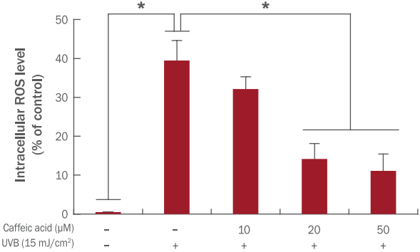
UVB는 여러 세포 내에서 ROS 생산에 관여하는 주요한 유도자 중 하나이다. 따라서, 본 연구에서는 카페인산의 UVB에 의한 세포독성 보호 효능이 ROS 생성 억제와 관련 있는지 파악하고자 하였다. 먼저 카페인산을 UVB 조사 전에 12 h 전처리 해주었다. UVB 조사 이후 24 h 배양시키고, 세포 내 존재하는 ROS level를 DCFDA fluorescent dye를 이용하여 분석하였다. 그 결과, UVB 조사는 모유두 세포 내 ROS level을 39.48±5.14% 증가시켰으나, 10, 20, 50 µM 카페인산을 전처리한 실험군에서는 UVB 조사 이후 세포 내 ROS level이 32.14±3.17%, 14.14±3.96%, 11.10±4.31%로 현저하게 감소하였다(Figure 3). 이러한 결과를 토대로, 진피 모유두 세포에 있어 카페인산에 의한 UVB 의존적 세포 손상 보호 효능은 ROS 생성억제 효능과 연관되어 있음을 나타낸다.

Caffeic acid downregulates the level of intracellular ROS in HFDPCs.
HFDPCs were treated with the indicated doses of caffeic acid for 12 h followed by irradiation to UVB. And then intracellular reactive oxygen species (ROS) levels were measured by using a flow cytometric analysis. The results are presented as M±S.D. of three independent experiments. * p<0.05 compared with control sample (untreated group of caffeic acid and UVB).
4. 인간 진피 모유두 세포에서 카페인산의 UVB에 의한 세포노화 보호 효과
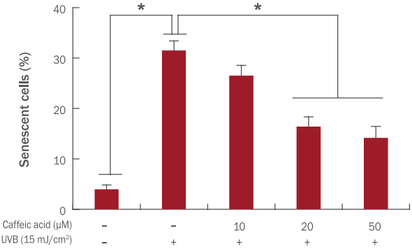
높은 수준의 ROS는 세포노화의 주요한 원인 중 하나로 알려져 있다. 기존 보고에서, 초기 탈모에서 나타나는 모유두 세포의 조기 노화(premature senescence)는 세포 내 SA-β-galactosidase의 발현 변화와 연관되어 있으며, 이러한 모유두 세포의 조기노화에는 산화적 스트레스가 관여되어 있을 것으로 보고 있다. 따라서, 본 논문에서는 추가적으로 자외선에 의한 모유두 세포의 세포노화 유발에 있어 카페인산의 효능을 분석하고자 하였다. 모유두 세포를 앞선 실험(Figure 3)과 동일하게 카페인산을 전처리한 후 UVB를 조사하여 나타나는 세포노화를 SA-β-galactosidase detection assay를 이용하여 분석하였다. UVB 조사는 대조군 대비 31.50±1.90% 세포노화가 증가됨을 확인하였다(Figure 4). 흥미롭게도, 자외선을 조사하기 전에 카페인산을 10, 20, 50 µM 농도로 전처리한 실험군에서는 세포노화의 비율이 각각 26.50±2.03%, 16.41±1.93%, 13.94±2.31%로 나타났다. 이를 통해, 카페인산은 UVB에 의한 모유두 세포의 세포노화를 현저하게 억제하는 효능이 있음을 밝혔다.

Caffeic acid inhibits cellular senescence induced by UVB irradiation in HFDPCs.
HFDPCs were treated with the indicated doses of caffeic acid for 12 h followed by irradiation to UVB. And then cellular senescence level was measured by using a senescenceassociated β-galactosidase assay. The results are presented as M±S.D. of three independent experiments. * p<0.05 compared with control sample (untreated group of caffeic acid and UVB).
Conclusion
본 논문에서는, UVB 의존적 모유두 세포 손상에 대한 카페인산의 보호 효능을 생화학적 분석법을 통해 증명하였다. 특히, 카페인산의 전처리는 UVB 조사에 의한 세포독성, 세포 내 ROS 생산, 세포노화를 현저하게 억제하였다. 비록 이러한 카페인산의 보호 효능 관련 특이적 세포신호전달체계를 증명하지 못하였으나, 본 연구논문은 탈모를 예방 및 억제할 수 있는 가능성 높은 물질로서 카페인산 발견에 그 중요성이 있다.
최근의 연구논문들은 탈모에 있어 산화적 스트레스에 대한 영향을 제시하고 있으며, 이는 산화적 스트레스의 억제는 탈모 및 백모화를 조절하는데 중요한 신규 전략으로 제시될 수 있음을 나타낸다(Trüeb, 2009). 과거 사례연구에 따르면, 탈모가 없는 정상인과 비교하여 원형 탈모(alopecia areata)를 가진 환자에서 산화적 스트레스가 상대적으로 높다는 것을 증명하였다(Bakry et al., 2014). 게다가, 마우스를 이용한 실험을 통해 염색약에 의해 유도되는 탈모는 대부분 hydrogen peroxide에 의해 유도되는 산화적 스트레스가 원인이라고 보고되었다(Seo et al., 2012).
산화적 스트레스는 모낭 성장 저해제 및 남성형 탈모(androgenic alopecia)의 주요한 원인으로 알려진 transforming growth factor β의 생산을 촉진시킨다(Lee & Hwang, 2009; Shin et al., 2013). 이전의 논문에서 카페인산은 인간 각질세포에 있어 UVA에 대한 광보호 효과(photoprotective effects)를 증명하였으며(Pluemsamran et al., 2012), 여러 임상 및 이론 논문들은 자외선은 산화적 스트레스, 휴지기 탈모(telogen effluvium), 모낭-미세염증(follicular micro-inflammation) 등의 유도를 통해 모발의 성장에 부정적인 영향을 나타냄을 밝혔다(Camacho et al., 1996; Johnsson et al., 1987; Trüeb, 2003).
따라서, 산화적 스트레스를 억제하는 것은 스트레스 및 안드로겐 의존적 탈모를 극복하는데 중요한 전략으로 사료될 수 있다. 본 연구에서는 카페인산은 모유두 세포에서 산화적 스트레스에 의한 세포 손상을 억제할 뿐만 아니라 모유두 세포에 세포독성을 거의 유발하지 않음을 확인하였다. 따라서, 추후 임상학적인 연구를 통해 카페인산의 두피 도포에 따른 영향을 분석함으로써, 카페인산을 탈모 예방 및 탈모억제의 기능성 물질로 개발하는데 기초 자료로서 활용될 수 있을 것으로 사료된다.
Acknowledgements
이 논문은 2016년도 건국대학교 KU학술연구비 지원에 의한 논문임.