


Introduction
Skin aging is a multifaceted process influenced by intrinsic and extrinsic factors, including genetics and environmental exposure (Papakonstantinou et al., 2012). It leads to the development of wrinkles, loss of elasticity, and thinning of the skin, affecting a person's social life (Longo, 2016). The cosmetics industry is continuously explores active ingredients to prevent and reduce skin aging, with cosmeceuticals playing a vital role in anti-aging skin care (Martin & Glaser, 2011). Besides addressing the signs of aging, cosmeceutical products should also promote "healthy aging" by providing psychological tranquility and alleviating stress (Ganceviciene et al., 2012). Chronic psychological stress accelerates skin aging by disrupting the skin barrier, activating the hypothalamic-pituitary-adrenal axis, and increasing reactive oxygen species (ROS) production (Dunn & Koo, 2013).
Moreover, the aging process is linked to the activation of inflammatory pathways in the skin, which is commonly referred to as "inflammaging" (Neves & Sousa-Victor, 2020). UVB radiation plays a significant role in skin damage and aging by stimulating cytokines and increasing the production of ROS (Abbas et al., 2018; Lago & Puzzi, 2019). Key cytokines such as IL-1β, IL-6, and TNF-α known to induce chronic inflammation and initiate the breakdown of collagen by upregulating MMP (Borg et al., 2013). Another major player in cellular inflammation, NF-κB, is upregulated by UV irradiation, resulting in chronic inflammation and photogenic reactions (Ansary et al., 2021; Pittayapruek et al., 2016). TNF-α, which plays a crucial role in skin inflammation, can cause irreversible damage to the epidermis (Bashir et al., 2009; Mirastschijski et al., 2019). Excessive production of IL-6, TNF-α, and IL-1 has been associated with skin damage and an increased risk of melanoma (Tam & Stepień, 2011).
Skin aging is a result of gradual depletion of collagen and elastin in the dermal connective tissue, leading to thinner skin with reduced mechanical strength and increase in the formation of wrinkles (Parente et al., 2015; Varani et al., 2001). The degradation of collagen and elastin primarily occurs due to the activity of matrix metalloproteinases (MMPs), which are zinc-containing proteinases capable of breaking down various extracellular matrix proteins (Nelson et al., 2000; Wang et al., 2008). Exposure to UV radiation can significantly increase levels of multiple MMPs, including MMP-1, MMP-3, and MMP-9, which are highly regulated by AP-1 (Quan et al., 2009). These MMPs possess the ability to break down collagen fibers, thereby contributing to the formation of wrinkles and tissue damage during the aging process of the skin (Pittayapruek et al., 2016; Shen et al., 2019).
Botanical ingredients and their derived products are widely utilized as major cosmeceutical agents in cosmetics due to the increasing awareness of health benefits and the preference for environmentally friendly consumer behavior (Amberg & Fogarassy, 2019; Kang et al., 2021; Sharmeen et al., 2021). Essential oils, which are complex mixtures of lipophilic plant metabolites with a molecular weight below 300 atomic mass units, exhibit beneficial bioactive properties owing to their low molecular weight and lipophilicity (Arena et al., 2021; Kazemi et al., 2020; Modarresi et al., 2019). These oils are extracted from various plant parts, such as leaves, flowers, barks, rhizomes, roots, and citrus fruit peels (Miguel, 2010; Je et al., 2021), and find applications in aromatherapy, pharmaceuticals, food, and clinical medicine industries (Firenzuoli et al., 2014). Inhaling or topically applying essential oils can restore mental and physical balance, alleviate stress, and provide skin care benefits (Aburjai & Natsheh, 2003; Ali et al., 2015; Jung & Lee, 2022). Additionally, essential oils serve as fragrances, preservatives, and active agents in cosmetics, apart from their pleasant aroma (Burnett et al., 2019; Sharmeen et al., 2021). Citrus essential oils, extracted from citrus fruit peels are classified into sweet and sour varieties, with the sour citrus being preferred in cosmetics due to its refreshing scent (Fukumoto et al., 2008; Nguyen & Sawamura, 2008). The volatile nature of essential oils enables them to interact with receptors in the central nervous system, influencing memory and mood through inhalation (Buchbauer & Jirovetz, 1994; Hongratanaworakit, 2004).
Citrus junos Siebold ex Tanaka (C. junos) is a widely cultivated plant in Korea, China, and Japan, known for its fragrant fruit used in Korean cuisine and traditional medicine (Kim et al., 2010). Extracts obtained from various parts of the fruit C. junos, such as the peel, seeds, and pulp, possess diverse chemical compositions and exhibit a range of biological activities (Song et al., 2019).
Extracts derived from C. junos have been found to possess anti-inflammatory, antioxidant, and anti-obesity effects (Shimada, 2015; Zang et al., 2014). Major constituents of C. junos extracts, such as naringin and hesperidin, have demonstrated anti-inflammatory properties in animal models (Nie et al., 2012). The extract from the peel scavenges free radicals and reduces nitric oxide production, while C. junos seed oil, containing unsaturated fatty acids, exhibits anti-inflammatory and antioxidant properties (Hong et al., 2017; Ko et al., 2020; Shin et al., 2010).
Moreover, limonoids derived from C. junos seeds have been shown to have a positive impact on beneficial intestinal microbiota (Minamisawa et al., 2021).
Additionally, Citrus junos essential oil (CJEO) with its unique aroma finds applications in the food, aromatherapy, and cosmetics industries. Inhalation of CJEO has demonstrated effects such as reduced heart rate, increased cerebral blood flow, and sedative and anxiolytic-like effects. CJEO also exhibits antimicrobial properties (El-Toumy & Hussein, 2020; Matsumoto et al., 2014; Satou et al., 2012).
The objective of this study is to evaluate the effects of CJEO on skin anti-aging and establish baseline data for future investigations. The chemical composition of CJEO was determined using gas chromatography-mass spectrometry (GC-MS). Its impact on cell viability, inflammation, collagen degradation, and collagen synthesis were assessed in HEKa and HDF cell lines. Quantitative RT-PCR was employed to measure mRNA levels and identify biomarkers associated with inflammation and skin elasticity in the aging process. Additionally, a quantitative analysis using HPLC was conducted to optimize the formulation with high concentrations of active CJEO ingredients. In vitro efficacy tests were followed by clinical studies assessing CJEO's effectiveness in improving elasticity. Safety evaluations were performed during and after CJEO emulsion application.
Materials and Methods
1. Preparation of CJEO
In 2020, C. junos fruits were sourced from Hansung Food Co., Ltd. (Korea). The fruits underwent authentication and were confirmed to be C. junos species. The essential oil was extracted from the fruit peels during the process of pressing the juice from the fruits. Subsequently, the obtained CJEO was stored under refrigeration at 4℃ to ensure sample preservation until the analyses were conducted. For the in vitro efficacy tests, CJEO was dissolved in a 10% dimethyl sulfoxide (DMSO) solution, followed by sonication for 5 minutes to facilitate proper mixing.
2. Cell lines and culture
Human dermal fibroblasts (HDF) were cultured in Fibroblast Basal Medium (FBM 106) supplemented with low serum growth supplement (LSGS), penicillin (100 IU/mL) and streptomycin (100 μg/mL). Human epidermal keratinocytes (HEKs) were cultured in Dulbecco's modified Eagle's medium (DMEM) supplemented with penicillin (100 IU/mL) and streptomycin (100 μg/mL), and 10% fetal bovine serum (FBS). Both HDF and HEKs cell lines were maintained in a 5% CO2 incubator at 37℃. Upon reaching confluency, the HDF and HEKa monolayer cultures were detached using 0.25% trypsin-EDTA solution.
3. Chemical composition analysis
The chemical composition of CJEO was analyzed by GC-MS using a Shimadzu TQ-8050NX instrument equipped with a SH-Rxi-5Sil MS column (30 m×0.25 mm, 0.25 μm film). The GC-MS analysis involved the following protocol: and initial column and oven temperature of 50℃, which was gradually increased to 250℃ and held for 3 to 5 minutes. The ion source temperature was set at 250℃, and real-time analytics software was employed. Prior to analysis, the CJEO sample was diluted in CHCl3 and filtered through a 0.45 μm PVDF membrane. A splitless injection of the diluted sample (1.0 μL) was used for analysis. Helium was used as the carrier gas at a flow rate of 1.00 mL/min. Component identification was based on retention indices relative to a homologous series of n-alkanes and mass spectral fragmentation patterns, which were compared with existing literature and an internal MS library. The relative amounts of components were calculated based on GC peak areas using response factors. Detailed analytical conditions for GC/MS can be found detailed in Table 1.
4. Cell viability assay
The viability of HEKs and HDF cells was assessed using a modified Mosmann's method based on the conversion of 3-(4,5-demethyl-2-thiazolyl)-2,5-diphenyl-2H-tetrazolium bromide (MTT) into formazan through mitochondrial oxidation (Mosmann, 1983). First, 1×10⁵ cells/well were seeded in a 96- well plate and cultured for 18 hours. Then, CJEO dissolved in 10% DMSO and deionized water was added to the cells at concentrations of 0, 10, 50, 100, 500, and 1000 μg/mL. After 24 hours of incubation, the medium was removed, the cells were washed with PBS, and a new medium was added. The cells were then treated with 5 mg/mL MTT solution (Sigma Aldrich Corp., USA). Formazan crystals produced from MTT were solubilized in 150 μL DMSO and the absorbance of each well was measured at 570 nm using a microplate reader (Epoch2C, USA). The optical density of formazan in control cells without CJEO was taken as 100% viability.
5. NO concentration
The quantification of nitric oxide (NO) production was carried out using the Griess reagent assay (Invitrogen, USA). HEKs were cultured in a 96-well plate for 24 hours, followed by exposure to 200 mJ/cm2 of UVB radiation for 10 minutes. Subsequently, CJEO was added to the wells at concentrations of 0, 5, 10, 50, and 100 μg/mL. After a 48-hour incubation, the supernatants were collected for analysis of NO content. To determine the NO concentration, a 150 μL aliquot of Griess reagent, consisting of 1% sulfanilic acid and 0.25% naphthylethylenediamine dihydrochloride) was added to 20 μL of each sample following the manufacturer's instructions. The absorbance of the resulting mixture was measured at 548 nm using a microplate reader. The NO concentration was calculated by referencing the nitrite standard curve and determining the nitrite concentration corresponding to the mean absorbance value.
6. RNA isolation and quantitative RT-PCR
1) The expression analysis of IL-1β, IL-6, NF-κB, and TNF-α
To analyze the expression of IL-1β, IL-6, NF-κB, and TNF-α, the following steps were followed. HEKs cells were incubated in DMEM supplemented with 10% FBS, 100 IU/mL penicillin, 100 μg/mL streptomycin, and maintained in a 5% CO2 environment at 37℃ for 24 hours. Subsequently, the cells were treated with various concentrations of CJEO and cultured for an additional 48 hours. Total RNA was isolated from the cells, and cDNA synthesis was performed using a PrimeScript 1st cDNA Synthesis kit. Amplification was carried out through 35 cycles using a Thermalcycler. The PCR products were visualized on a 1% agarose gel, and the relative gene expression was analyzed using a gel documentation system.
2) The expression analysis of MMP-1, MMP-3, COL1A1, COL3A1
To determine the mRNA expression of MMP-1, MMP-3, COL1A1, and COL3A1, HDF cells were cultured in Medium 106 supplemented with 1×LSGS, 100 IU/mL penicillin and 100 μg/mL streptomycin in a 5% CO2 environment at 37℃ for 24 hours. After removing the medium the fibroblasts were treated with various concentrations of CJEO (5, 10, 50, 100 μg/mL) and cultured for 48 hours. Total RNAs from each well were isolated using the TransZol reagent. The obtained total RNA was used to generate the cDNA using the PrimeScript 1st cDNA Synthesis kit, following the manufacturer's instructions. PCR amplifications were performed in a thermalcycler with 35 cycles: denaturation at 95℃ for 1 minute, annealing at 50 to 60℃ for 30 seconds, and extension at 72℃ for 1 minute. The primer sequences and further details of PCR are provided in Table 2. GAPDH served as a control gene. PCR products were electrophoresed on a 1% agarose gel and visualized using ethidium bromide staining. A gel documentation system was utilized to analyze the results and determine the relative ratio of changes in the target genes compared to the control.
7. Clinical studies
The study protocol and the informed consent form were approved by an independent regional ethical committee (DrSSL-2021-56; Dermacosmetics Institutional Biosafety and Ethics Committee, Korea), and written informed consent was obtained from all participants before their participation in this research. This study included only female participants, with a total of 20 to 60 years of age (mean age 39.1 years), who were recruited to ensure a comprehensive evaluation. An oil-in-water (O/W) emulsion was meticulously developed, incorporating specially selected ingredients to enhance and maintain skin homeostasis. The emulsion formulation comprised CJEO, identified as the O/W emulsion type with the highest CJEO concentration determined in the previous HPLC quantification study. In addition, a placebo emulsion without CJEO was also formulated.
For dermal patch testing, three emulsions with different concentrations of CJEO were prepared. In the primary skin irritation patch test, each formulation was individually applied to participants' skin. The test products included the placebo emulsion (without CJEO), 0.15% CJEO emulsion, and 0.3% CJEO emulsion. All formulations were prepared under similar conditions as outlined in Table 3.
Based on the results of the dermal patch tests, the placebo emulsion and 0.3% CJEO emulsion, both demonstrating no skin irritation, were selected for the subsequent 4-week clinical trial.
The clinical trial consisted of three visits: an initial screening, scoring, and evaluation visit, followed by evaluations at week 2 and week 4. After obtaining informed consent and medical history, participants underwent screening for inclusion and exclusion criteria. Random assignment was conducted, with participants being assigned to either the control group (placebo emulsion) or the experimental group (CJEO emulsion). Blinding of the test materials was ensured. Participants were instructed to apply the test products to their inner arms, using gentle circular motions, every morning and night for 4 weeks after thoroughly washing and drying their hands. The test area remained free of other topical treatments.
During each visit, tolerance and elasticity evaluations were conducted in the same designated areas. Skin elasticity was objectively measured using a Cutometer dual MPA 580. The efficacy of the test products was assessed based on elasticity measurements (E, mm) after 2 weeks and 4 weeks. The condition of the skin and evaluation parameters were recorded, and any adverse events were carefully documented throughout the study.
8. Statistical analysis
For the in vitro experiments, mean±standard deviation (SD) was presented based on at least three independent experiments. Data analysis employed the student's t-test using Microsoft Excel 365 (Microsoft, USA). A significance level of p<0.05 was deemed indicative of statistical significance.
In the context of the clinical trial, a cohort of at least 20 participants was recruited. Rigorous statistical analysis was applied to assess the impact of the tested products on pre- and post-product usage data, with significance set at p<0.05. To validate the statistical analyses, tests were performed to confirm the assumptions of normality and sphericity. This encompassed the use of both parametric methods, such as Two-way Repeated Measures ANOVA and Two-way Repeated Measure MANOVA, as well as a non-parametric approach utilizing the LD.F2 test.
Additionally, post-hoc analyses were conducted, employing the Bonferroni correction method to adjust for multiple comparisons. These meticulous statistical techniques were thoughtfully chosen to ensure the robustness and reliability of our findings, adhering to the highest scientific standards.
Results
1. Chemical Composition of CJEO
The chemical composition of CJEO was analyzed using gas chromatography-mass spectrometry (GC-MS), which identified a total of 26 volatile components. These components were classified into 7 oxygenated terpenes and 19 non-oxygenated terpenes, with 12 being monoterpenes and 7 being sesquiterpenes. Among the identified constituents, the major compounds in CJEO were α-pinene, β-myrcene, p-cymene, d-limonene, γ-terpinene, linalool, and β-farnesene. The relative proportions of each component were determined based on GC peak area. The quantitative data for the major components is presented in Table 4.
2. Effect of CJEO on cell viability
To assess the impact of CJEO on cell viability, the MTT assay was performed on HEKa and HDF cell lines. The results showed a dose-dependent decrease in cell viability with increasing concentrations of CJEO across all three cell lines. Particularly, the most significant reduction in cell viability was observed at a concentration of 500 μg/mL of CJEO. For subsequent experiments, a concentration of 100 μg/mL of CJEO, which exhibited approximately 90% cell viability, was selected.
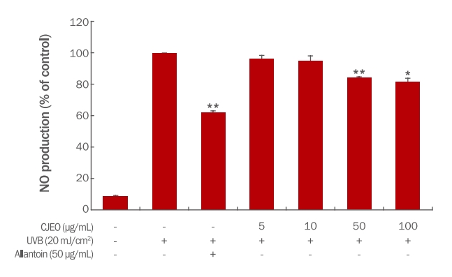
3. Inhibition of nitric oxide (NO) production
The inhibitory effect of CJEO on nitric oxide (NO) production was evaluated in HEKa cells. Treatment with CJEO lead to a notable reduction in NO production compared to the untreated control group. While all tested concentrations of CJEO displayed a decrease in NO production, the inhibitory effect was relatively weaker than that of the positive control, allantoin (Figure 1). These findings suggest that CJEO possesses the potential to suppress NO production in HEKa cells, indicating its anti-inflammatory properties.
4. Effects of CJEO on gene expression
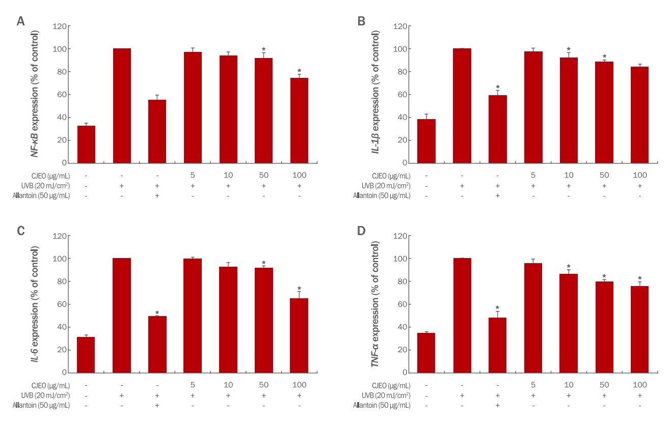
1) Expressions of TNF-α, IL-1β, IL-6, and NF-κB
To evaluate the impact of CJEO on mRNA expression in UV-induced HEKs cells, the expression levels of TNF-α, IL-1β, IL-6, and NF-κB were measured using quantitative RT-PCR (qRT-PCR). The results showed that CJEO significantly inhibited the expressions of TNF-α, IL-1β, IL-6, and NF-κB in a dosedependent manner (p<0.05). However, it is important to note that the anti-inflammatory activity of CJEO was relatively weaker when compared to the 50 μg/mL concentration of allantoin, as depicted in Figure 2. Nevertheless, these findings demonstrate that CJEO treatment effectively reduces the expression of inflammatory biomarkers in a dose-dependent manner.
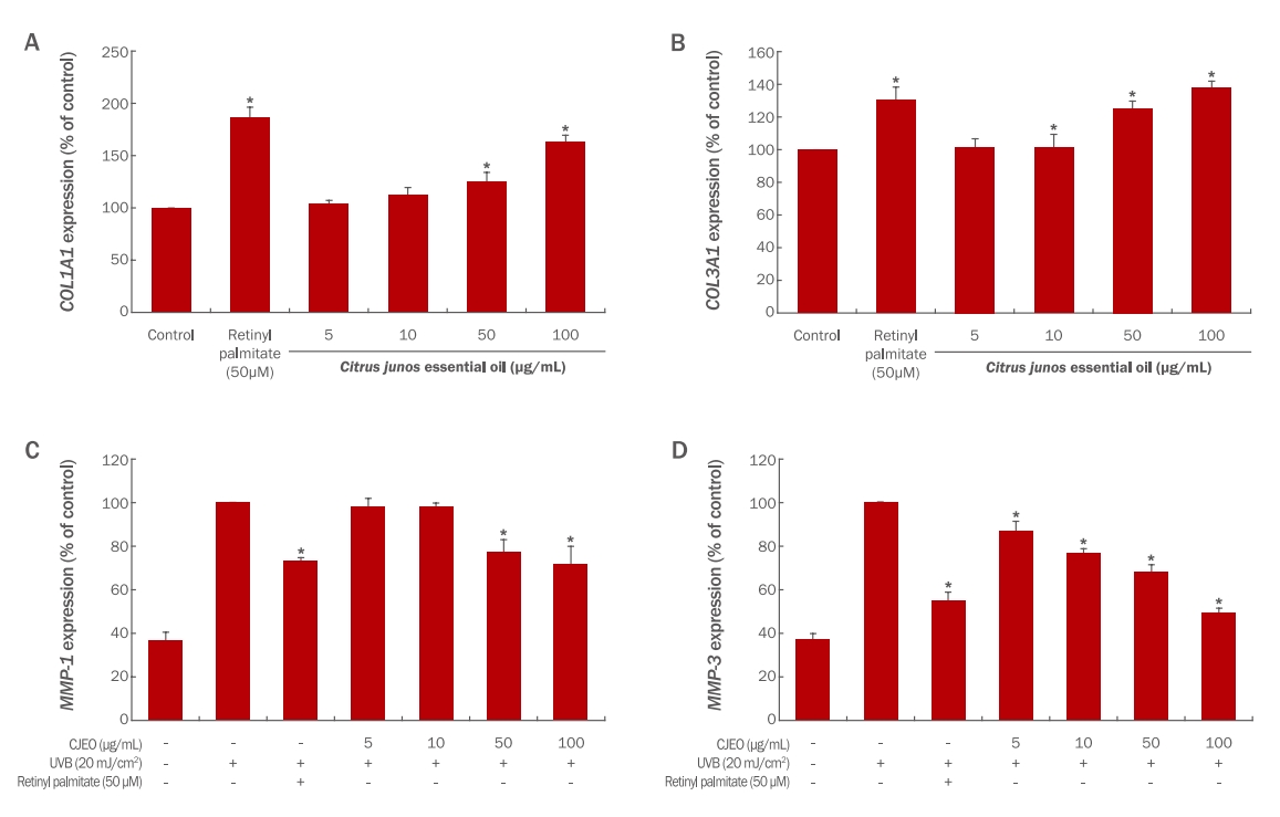
2) Expressions of MMP1, MMP3, COL1A1, and COL3A1
To determine the impact of CJEO on mRNA expression in HDF cells, the expressions of collagen degradation-related genes, MMP1 and MMP3, were assessed using quantitative RT-PCR (qRT-PCR).
The results demonstrate that CJEO dose-dependently inhibits the expression of MMP1 and MMP3, as illustrated in Figure 3. At a concentration of 100 μM/mL, CJEO exhibited greater inhibitory effects on MMP1 and MMP3 expression compared to the 50 μM concentration of retinyl palmitate.
Furthermore, the expressions of collagen synthesis-related genes, COL1A1 and COL3A1, were also assessed using quantitative RT-PCR (qRT-PCR) in HDFs.
Treatment with CJEO at concentrations of 50, and 100 μg/mL resulted in a significant increase in COL1A1 expression to 125.56%, and 163.67%, respectively (p<0.05). The positive control treatment with retinyl palmitate (50 μM) significantly increased COL1A1 expression to 186.37%, as depicted in Figure 3.
Similarly, treatment with CJEO at concentrations of 10, 50, and 100 μg/mL resulted in a significant increase in COL3A1 expression to 101.47%, 125.22%, and 137.89%, respectively (p<0.05). The positive control also significantly increased COL3A1 expression to 130.44%, as shown in Figure 3.
Overall, the expressions of COL1A1 and COL3A1 were significantly increased by CJEO in a dose-dependent manner (p<0.05).
5. Dermal patch test
A dermal patch test was conducted using an IQ Ultimate patch loaded with 20 μL of CJEO emulsion at two different concentrations (0.15 and 0.3%). The patches were applied to the backs of 30 participants for 24 hours. Skin reactions were assessed by a single trained assessor at 1 and 24 hours after patch removal, using modified York's methods (York et al., 1995). The assessment included evaluation erythema, spots, edema, and vesicles. Each skin reaction was given a score ranging from 0 to 4, where a score of "0" indicated no visible reaction, and higher scores represented increasing severity of erythema, edema, and vesicular erosion. The scoring was done on a simple scale, e.g. 0, +, ++, +++, ++++ as described in Table 5.
The dermal irritation index was calculated using the following equation.
The mean dermal irritation index was categorized into the following levels: No irritation for scores between 0.00 and 0.25, slight irritation for scores between 0.26 and 1.00, moderate irritation for scores between 1.01 and 2.50, and severe irritation for scores between 2.51 and 4.00. In this study, all participants who underwent the dermal irritation index assessment at the concentrations of 0.15% and 0.3% of CJEO obtained a dermal irritation index of 0.00. These consistent results from the two assessments for primary skin irritation, across all participants, provide compelling evidence suggesting a lack of significant irritation potential associated with the CJEO emulsion at these concentrations.
6. Skin elasticity assessment
After randomly allocation of participants' left and right anterior forearms into the control and experimental groups, both groups used their respective products for 4 weeks, after which skin elasticity (E, mm) was measured. As shown in Table 6, the experimental group exhibited a significantly greater improvement in skin elasticity (E, mm) compared to the control group. These findings are consistent with the observations made after randomly allocating participants' left and right anterior forearms into the control and experimental groups, with both groups using their respective products for 4 weeks before measuring skin elasticity (E, mm). The statistical significance of this difference was confirmed (p<0.05), providing comprehensive insights into the efficacy of the experimental product.
7. Safety evaluation
During the study, participants did not report any discomfort during the application or post-application period. Additionally, no persistent erythema or post-inflammatory hyperpigmentation was observed. The CJEO emulsion was well-tolerated, and no adverse effects were reported throughout the study.
Conclusions
The exploration into the biological efficacy of CJEO as a bioactive ingredient for anti-aging products divulged its potent anti-aging capabilities through an integrative approach encompassing chemical composition analysis, in vitro experimentation, and clinical trials. The chemical composition analysis through GC-MS revealed 26 volatile components within CJEO, categorized into 7 oxygenated terpenes and 19 non-oxygenated terpenes, including 12 monoterpenes and 7 sesquiterpenes. Noteworthy compounds encompassed α-pinene, β-myrcene, p-cymene, d-limonene, γ-terpinene, linalool, and β-farnesene.
CJEO exhibited notable inhibition of NO production, and qRT-PCR analysis illuminated its potential in mitigating skin inflammation through decreased pro-inflammatory cytokines and augmented collagen synthesis markers, indicative of anti-inflammatory and skin-firming properties. However, to deepen comprehension, further investigations should delve into each specific constituent's effects and potential synergistic interactions.
Furthermore, clinical trials underscored CJEO emulsion's efficacy in enhancing skin elasticity. While both CJEO and placebo emulsions boosted skin elasticity, the former showcased significant advancement from weeks 2 to 4 post-application, in contrast to the placebo group. Despite debates around limonene, a major CJEO component, pertaining to possible skin irritation, our clinical trial established the CJEO emulsion's safety through skin irritation tests and comprehensive assessments. In essence, this study confirms CJEO's prowess as an anti-aging ingredient.
This work is part of Byel Kim's Ph.D. thesis at Konkuk University, Seoul, Korea.